
Page 97 - IJB-8-3
P. 97
Hu, et al.
On the 4 day after surgery, the patient developed internal fixation, and autogenous iliac bone grafting for
th
a fever up to 38.8℃. Wound drainage significantly occipitocervical fusion (Plan B).
increased, which was thought to be a result of either An anterior cervical approach was used to resect
cerebrospinal fluid leakage or infection. Bacterial culture C2 along with the tumor, the C1 anterior arch and the
of the fluid showed the presence of Gram-positive coccus. C2-3 intervertebral disc. However, after placing the
Based on susceptibilities, vancomycin was given and 3D-printed artificial vertebra into the resulting bone
was able to quickly control the infection. The infection defect, we found that the prosthesis was smaller than
had not yet caused any adverse effects on the artificial the body’s actual bone defect, resulting in a lack of
vertebral body implant, therefore the decision was made fixation. We then converted to a conventional spinal
to retain the hardware. She experienced a lengthy hospital reconstruction (Plan B). Post-operative pathology
stay (22 days). During the 13-month-follow-up after the results confirmed osteosarcoma. Post-operative X-ray
surgery, the patient moved freely and had no pain and or showed excellent reconstruction, according to Plan B
local recurrence. (Figure 9E). At the 37 month after surgery, the patient’s
th
neck moved freely but with pain. He was diagnosed with
3.3. Unsuccessful implantation osteosarcoma local recurrence and was recommended to
(1) Case 7 receive chemo-radiotherapy.
A 51-year-old male visited FUSCC for neck pain, (2) Case 8
limitation of motion, and numbness of both upper A 42-year-old male underwent L1 tumor resection
extremities. Pre-operative X-ray and CT showed a C1 in another hospital 4 years ago. He was admitted to
and C2 tumor resulting in pathological fracture and FUSCC for lower back pain and activity limitation.
dislocation (Figure 9A). The tumor was located in X-ray revealed failed hardware, with a fractured rod and
C1 ~ 2 (Figure 9B). We planned to en bloc resect C2, L1 artificial vertebral body subsidence (Figure 10A).
partially resect and correct alignment of C1, and design We planned to design a 3D-printed vertebral body to
a customized 3D-printed artificial vertebral body to reconstruct his spine. CT-derived 3D imaging visually
reconstruct his spine (Plan A) (Figure 9C). Final titanium displayed the lesion (Figure 10B). Individualized
alloy implants were manufactured (Figure 9D). An 3D-printed artificial vertebral body and screw rod
alternative preoperative traditional spinal reconstruction internal fixation system were designed and modeled
plan was prepared, including bone cement formation,
(Figure 10C). During the operation, T12 and L2 bone
graft beds were polished. However, the 3D-printed
A B prosthesis could not be completely implanted because
of mismatch between the 3D-printed prosthesis and the
bone defect height. An alternative solution that allows
for extendable artificial vertebra with incorporation of
autogenous rib and allogeneic bone was performed on
the patient (Figure 10D). During the 4-month-follow-
up after the surgery, the patient moved freely and had
no pain.
C D 3.4. Surgical characteristics
Of the eight patients, six succeeded in 3D-printed spinal
implantation, two failed and converted to conventional
reconstruction. For patients with 3D-printed spinal
implants: (i) the median surgery time was 414 min; (ii)
the median blood loss was 2,150 ml; (iii) the median
blood transfusion was 2000 ml; (iv) the median length
of hospital stay was 9 days; (v) four underwent adjuvant
therapy after the surgery; and (vi) they experienced no
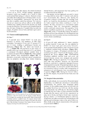
Figure 8. Case 6. (A) Pre-operative X-ray and MRI showing tumor pain, moved freely, and had no local tumor recurrence
recurrence at the T2~4 levels; (B) 3D imaging visually showing
T2~4, planned for resection and replacement with patient-specific during a median 11.5 months-post-operative follow-up.
3D-printed artificial vertebral body; (C) surgical clinical photo All patients had stable reconstructions without failure and
demonstrating the autogenous bone granule used within the porous kept in good performance status at the end of follow-up.
part of the prosthesis to promote bone fusion; (D) post-operative More details on the clinical characteristics of the patients
imaging showing well positioned implant. are presented in Table 2.
International Journal of Bioprinting (2022)–Volume 8, Issue 3 89