
Page 92 - IJB-8-3
P. 92
3D-Printed Artificial Vertebral Body
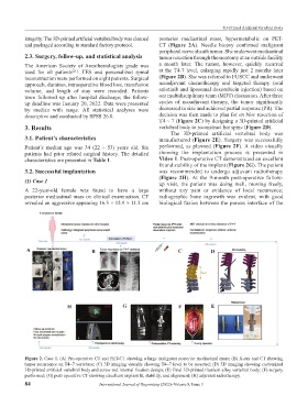
integrity. The 3D-printed artificial vertebral body was cleaned posterior mediastinal mass, hypermetabolic on PET-
and packaged according to standard factory protocol. CT (Figure 2A). Needle biopsy confirmed malignant
peripheral nerve sheath tumor. She underwent mediastinal
2.3. Surgery, follow-up, and statistical analysis tumor resection through thoracotomy at an outside facility
The American Society of Anesthesiologists grade was a month later. The tumor, however, quickly recurred
used for all patients . TES and personalized spinal at the T4-7 level, enlarging rapidly just 2 months later
[11]
reconstruction were performed on eight patients. Surgical (Figure 2B). She was referred to FUSCC and underwent
approach, duration, intraoperative blood loss, transfusion neoadjuvant chemotherapy and targeted therapy (oral
volume, and length of stay were recorded. Patients anlotinib and liposomal doxorubicin injection) based on
were followed up after hospital discharge; the follow- our multidisciplinary team (MDT) discussion. After three
up deadline was January 20, 2022. Data were presented cycles of neoadjuvant therapy, the tumor significantly
by median with range. All statistical analyses were decreased in size and achieved partial response (PR). The
descriptive and conducted by SPSS 26.0. decision was then made to plan for en bloc resection of
T4 ~ 7 (Figure 2C) by designing a 3D-printed artificial
3. Results vertebral body to reconstruct her spine (Figure 2D).
The 3D-printed artificial vertebral body was
3.1. Patient’s characteristics manufactured (Figure 2E). Surgery was successfully
Patient’s median age was 34 (22 – 53) years old. Six performed, as planned (Figure 2F). A video visually
patients had prior related surgical history. The detailed showing the implantation process is presented in
characteristics are presented in Table 1. Video 1. Post-operative CT demonstrated an excellent
fit and stability of the implant (Figure 2G). The patient
3.2. Successful implantation was recommended to undergo adjuvant radiotherapy
(Figure 2H). At the 9-month post-operative follow-
(1) Case 1
up visit, the patient was doing well, moving freely,
A 22-year-old female was found to have a large without any pain or evidence of local recurrence;
posterior mediastinal mass on clinical examination. CT radiographic bone ingrowth was evident, with good
revealed an aggressive appearing 16.3 × 15.9 × 11.3 cm biological fusion between the porous interface of the
A B C D
H G F E
Figure 2. Case 1. (A) Pre-operative CT and PET-CT showing a large malignant posterior mediastinal mass; (B) X-ray and CT showing
tumor recurrence on T4~7 vertebrae; (C) 3D imaging visually showing T4~7 level to be resected; (D) 3D imaging showing customized
3D-printed artificial vertebral body and screw rod internal fixation design; (E) Final 3D-printed titanium alloy vertebral body; (F) surgery
performed; (G) post-operative CT showing excellent implant fit, stability, and alignment; (H) adjuvant radiotherapy.
84 International Journal of Bioprinting (2022)–Volume 8, Issue 3