
Page 214 - IJB-8-4
P. 214
3D Printed Porous 45S5 Bioceramic
interconnection, and mechanical properties [8,9] . In recent fluidity, hindering its applications [20] . Hence, it is very
years, various artificial bone scaffold materials, such as important to adjust the process parameters.
metal [10] , bioceramic [11,12] , polymer [13] , and other kinds In this study, a low-cost LCD strategy was used
of composites [14,15] , have undergone rapid development to print porous 45S5 bioglass bone scaffolds. Different
and been successfully used as bone substitutes. aspects from design to printing process, including
However, the precise size of the bone scaffolds requires percentage content of dispersant, reheated temperature,
specific design. Based on these issues, 3D printed and mixed ratio of 45S5 suspension, were studied.
scaffolds have been extensively studied for customized Different structures and porosities that may affect the
bone tissue repair, which employs a combination of compressive strength of final scaffolds were investigated.
materials from computation 3D models and layer-by- X-ray crystallography (XRD), scanning electron
layer fabrication. Thus, many different structures have microscopy (SEM), micro-computed tomography (micro-
been designed [16,17] and various fabrication techniques CT), and mechanical compression test were used for
have been employed to produce porous scaffolds analyzing the sintered scaffolds. Our results demonstrated
through 3D printing technology, such as laser-based [18] , that the proposed 45S5 scaffolds fabricated by LCD mask
extrusion-based [19,20] and inkjet printing [21] , as well as stereolithography technology are well-designed in terms
lithography rapid prototyping [22] . Lithography-based of composition, morphology, porosity, and mechanical
3D printing such as digital light processing (DLP) and properties. This scaffold could be a promising substitute
liquid crystal display (LCD) may fabricate the bioglass in bone tissue repair.
or ceramic scaffolds using photopolymer and ceramics 2. Materials and methods
composite, and further sintering is needed to obtain
the scaffolds [23,24] . Hence, it is necessary to focus on 2.1. Design of 3D scaffolds and finite analysis
the shape and topology optimization to reduce cost
in the design of bone scaffold with good porosity and To build 3D scaffold models, Solidworks 2020 software
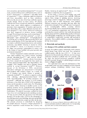
mechanical properties [25] . (Solid Works Corp, SUA) was used. Two cylindrical
Various biomedical ceramic materials used as the scaffold models with the size of Φ 15 × 15 mm were
bone repair scaffolds were widely studied, including designed, but the porosity was different: one was average
calcium phosphate-based [26,27] and calcium silicate- perforated cylindrical scaffold with 53% porosity, and
based bioceramics [28,29] . Calcium silicon bioceramics the other was layered perforated cylindrical scaffold
have been extensively investigated in bone repair due with 43% porosity. Images of scaffold design models are
to their good biocompatibility and degradability. For shown in Figure 1A and B.
example, 45S5 bioglass was used to prepare porous The equivalent stress and total deformation of the
scaffold in various biomedical fields [30,31] . However, model under axial load were evaluated by finite element
because of the brittleness of 45S5, conventional analysis software (NASDAQ: ANSS). Under linear
fabrication techniques are unsuccessful in fabricating
final products with complex shapes. As a result, the A B
use of bioglass was mostly limited to powder or
composite scaffold [32,33] . To achieve a superior scaffold
after sintering, photopolymerization-based LCD
mask stereolithography 3D printing technique was
proposed [34] . Although the accuracy of LCD printing
technique is less than that of DLP, LCD equipment is
cheap, and its operation is much easier. For 3D printing
of ceramic, further sintering is usually needed, so the C
printing accuracy can be comprehensively considered
during sintering even if LCD is less accurate than DLP.
Furthermore, 3D printed bioceramic scaffold for bone
tissue does not need high precision (printing accuracy
<50 um). In view of this, the specimens were fabricated
through LCD in this study. LCD has the advantages of
fast printing speed, good molding, and good printability
with higher concentration ceramic powders in the inks.
The porosity precision is controllable by designing Figure 1. (A) Average perforated cylindrical scaffold model. (B)
the sample structure. However, a higher proportion of Layered perforated cylindrical scaffold model. (C) Corresponding
ceramic suspension would increase the viscosity and equivalent stress by finite element analysis simulation.
206 International Journal of Bioprinting (2022)–Volume 8, Issue 4