
Page 219 - IJB-8-4
P. 219
Dong, et al.
A C
B
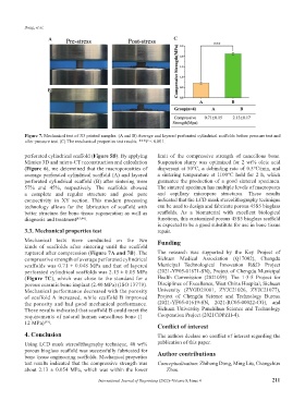
Figure 7. Mechanical test of 3D printed samples. (A and B) Average and layered perforated cylindrical scaffolds before pressure test and
after pressure test. (C) The mechanical properties test results. ***P < 0.001.
perforated cylindrical scaffold (Figure 5B). By applying limit of the compressive strength of cancellous bone.
Mimics 3D and micro-CT reconstruction and calculation Suspension slurry was optimized for 2 wt% oleic acid
(Figure 6), we determined that the macroporosities of dispersant at 50°C, a debinding rate of 0.5°C/min, and
average perforated cylindrical scaffold (A) and layered a sintering temperature at 1100°C hold for 2 h, which
perforated cylindrical scaffold (B) after sintering were guarantee the production of a good sintered specimen.
57% and 45%, respectively. The scaffolds showed The sintered specimen has multiple levels of macropores
a complete and regular structure and good pore and capillary micropore structures. These results
connectivity in XY section. This modern processing indicated that the LCD mask stereolithography technique
technology allows for the fabrication of scaffold with can be used to design and fabricate porous 45S5 bioglass
better structure for bone tissue regeneration as well as scaffolds. As a biomaterial with excellent biological
diagnosis and treatment [43,44] . functions, this customized porous 45S5 bioglass scaffold
is expected to be a good substitute for use in bone tissue
3.3. Mechanical properties test repair.
Mechanical tests were conducted on the two Funding
kinds of scaffolds after sintering until the scaffold
ruptured after compression (Figure 7A and 7B). The The research was supported by the Key Project of
compressive strength of average perforated cylindrical Sichuan Medical Association (Q17002), Chengdu
scaffolds was 0.71 ± 0.048 MPa and that of layered Municipal Technological Innovation R&D Project
perforated cylindrical scaffolds was 2.13 ± 0.05 MPa (2021-YF05-01871-SN), Project of Chengdu Municipal
(Figure 7C), which was close to the standard for a Health Commission (2021059). The 1·3·5 Project for
porous ceramic bone implant (2.40 MPa) (ISO 13779). Disciplines of Excellence, West China Hospital, Sichuan
Mechanical performance decreased with the porosity University (ZYGD21001, ZYJC21026, ZYJC21077),
of scaffold A increased, while scaffold B improved Project of Chengdu Science and Technology Bureau
the porosity and had good mechanical performance. (2021-YF05-01619-SN, 2021-RC05-00022-CG), and
These results indicated that scaffold B could meet the Sichuan University Panzhihua Science and Technology
requirements of natural human cancellous bone (1 – Cooperation Project (2021CDPZH-4).
12 MPa) [45] .
Conflict of interest
4. Conclusion The authors declare no conflict of interest regarding the
Using LCD mask stereolithography technique, 40 wt% publication of this paper.
porous bioglass scaffold was successfully fabricated for Author contributions
bone tissue engineering scaffolds. Mechanical properties
test results indicated that the compressive strength was Conceptualization: Zhihong Dong, Ming Liu, Changchun
about 2.13 ± 0.054 MPa, which was within the lower Zhou
International Journal of Bioprinting (2022)–Volume 8, Issue 4 211