
Page 274 - IJB-8-4
P. 274
Multifunctional 3D Bioprinting System for Construction of Complex Tissue Structure Scaffolds
The previous work has introduced the effect of UV light microextrusion (mesh and turtle models, Figure 7C)
irradiation on the biological activity of the cells in printed and pneumatic microextrusion (models of ear, mesh,
structures when GelMA is used as a bioink [61,62] . Therefore, and multilayer hydrogel skin-like structure, Figure 7D).
this section uses the previous experimental conclusions GelMA (10% [w/t]) was also used to print the structural
to directly verify the structure printing capability of the models of elephant head and spinal cord (Figure 8A). It can
system and the cell survival rate after printing. be seen from these results that the printing structure is well
According to the results of rheological analysis formed and has high stability and fidelity. The configured
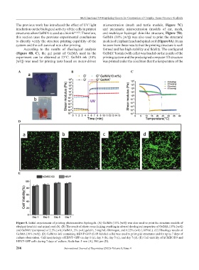
(Figure 8B, C), the gel point of GelMA used in the GelMA bioink (with cells) was loaded on the nozzle of the
+
experiment can be obtained at 23°C. GelMA ink (10% printing system and the predesigned computer 3D structure
[w/t]) was used for printing tests based on motor-driven was printed under the condition that the temperature of the
A B C
D
E
Figure 8. Initial experiments of printing photosensitive hydrogels. (A) GelMA (10% [w/t]) was also used to print the structure models of
elephant head (a) and spinal cord (b). (B) The result of photo cross-linking resulting in altered rheological properties of GelMA (10% [w/t])
and GelMA (composed of 2.5% [w/t] GelMA, 5% [w/t] gelatin, 5 mg/mL fibrinogen, and 0.25% (w/t) LAP/mL). (C) Rheology results of
+
GelMA (10% [w/t]). (D) GelMA+ ink containing HBVP-GFP (GFP-labeled cells) was used to print grid structures and for up to 7 days of
culture observation. Cell morphology of HBVP-GFP on day 1 (a), day 3 (b), day 5 (c), and day 7 (d). (E) Cell viability of hCMEC/D3 and
HBVP-GFP cells during 7 days of culture. Scale bar: 5 mm (A), 500 μm (D).
266 International Journal of Bioprinting (2022)–Volume 8, Issue 4