
Page 275 - IJB-8-4
P. 275
Xu, et al.
print head is 24°C (the needle size is 25 G, the movement diameter of 200 μm, the measured printing filament
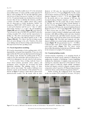
speed of the nozzle is 5 mm/s, and the ejection speed is 0.8 diameter is 197.11 ± 37.93 μm (Figure 9A). For the nozzle
mm /s). After printing, the 365 nm blue light that comes with an inner diameter of 300 μm, the measured printing
3
with the nozzle head was used to irradiate it at 5 mW/cm filament diameter is 295.67 ± 37.94 μm (Figure 9B).
2
for 30 s. The printed sample was incubated in an incubator For the nozzle with an inner diameter of 400 μm, the
for more than 20 min to stabilize the structure, and then, measured printing filament diameter is 432.00 ± 72.64
a picture was taken with a digital camera with a macro μm (Figure 9C). For the nozzle with an inner diameter
lens for observation or further incubation. GelMA ink of 600 μm, the measured printing filament diameter is
+
containing HBVP-GFP (GFP-labeled cells) was used 499.34 ± 110.85 μm (Figure 9D). For the nozzle with an
to print grid structures and for up to 7 days of culture inner diameter of 800 μm, the measured printing filament
observation. On days 1, 3, 5, and 7, the cell morphology diameter is 624.18 ± 114.35 μm (Figure 9E). On the basis
(Figure 8D) and cell viability (Figure 8E) were detected. of these process data, the ability to print complex PCL
The cell survival rate of hCMEC/D3 and HBVP cells after structures is further verified. Including square and circular
printing exceeded 80%, which met the requirements for structures with different filling styles and sizes, brackets
further use. The cells in the hydrogel began to stretch on for the femoral head, and tibial model structures were
the 3 day, and some cells joined together on the 7 day printed using a motor-driven piston-based microextrusion
th
rd
(Figure 8D[a-d]). This shows that the printing process nozzle (Figure 7E). Two-layer grid supports and cube-
does not cause damage to the cells. Therefore, the photo shaped scaffolds were printed using a pneumatic extrusion
cross-linking material-based printing nozzle developed in nozzle (Figure 7F, the gas pressure was 0.5 ~ 0.6 MPa).
this paper meets the basic printing needs. Cardiovascular stents were printed using a mechanical
screw-based nozzle (Figure 7G). The above results
3.3. Fused deposition modeling showed that this printing system has good forming quality
PCL has the characteristics of a low melting point (~60°C), in the formation of complex PCL structures.
degradability, and good biocompatibility. It has also been 3.4. Coaxial printing results
approved by the FDA and is an ideal material for biological
3D printing stents. Because the melting point of PCL is Coaxial extrusion bioprinting is an emerging branch of
relatively low, the temperature will soon drop below 37°C, bioprinting that shows strong potential in enhancing the
which is not damaging to the cells, after hot melt printing. engineering versatility of bioprinting. Coaxial bioprinting
In this system, three printing methods based on facilitates the fabrication of complex tissue structures by
motor driven pistons, pneumatic driven pistons, and enabling the concentric deposition of biomaterials. It was
mechanical screw extrusion are developed for fused initially applied to fabricate simple tubular vasculature
deposition modeling. The printing results of these but has now largely evolved to integrate complex cellular
three nozzles were tested. The results of different inner composition and self-assembly, ECM patterning structures,
diameter nozzles on the diameter of the printed PCL are controlled release, and multimaterial gradient profiles .
[63]
shown in Figure 9 (using a motor-driven piston-based Before printing, the configured bioink was loaded
microextrusion nozzle). For the nozzle with an inner on the two nozzles (30% [w/t] PF127 in inner, 10% [w/t]
A B C D E
Figure 9. The results of different inner diameter nozzles on the diameter of the printed PCL. Scale bar: 2 mm.
International Journal of Bioprinting (2022)–Volume 8, Issue 4 267