
Page 47 - IJB-9-1
P. 47
International Journal of Bioprinting Effect of ionic crosslinking on composite membranes
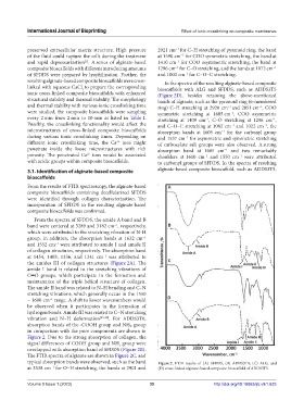
preserved extracellular matrix structure. High pressure 2921 cm for C–H stretching of pyranoid ring, the band
−1
of the fluid could rupture the cells during the treatment at 1590 cm for COO symmetric stretching, the band at
−1
and rapid depressurization . A series of alginate-based 1410 cm for COO asymmetric stretching, the band at
[1]
−1
composite bioscaffolds with different introducing amounts 1296 cm for C–O stretching, and the bands at 1072 cm
−1
−1
of SFDDS were prepared by lyophilization. Further, the and 1002 cm for C–O–C stretching.
−1
resulting alginate-based composite bioscaffolds were cross- In the spectra of the resulting alginate-based composite
linked with aqueous CaCl to prepare the corresponding bioscaffolds with ALG and SFDDS, such as ADDS3T5
2
ionic cross-linked composite bioscaffolds with enhanced (Figure 2D), besides retaining the above-mentioned
structural stability and thermal stability. The morphology bands of alginate, such as the pyranoid ring (6-membered
and thermal stability with various ionic crosslinking time ring) C–H stretching at 2920 cm and 2851 cm , COO
−1
−1
were studied, the composite bioscaffolds were sampling symmetric stretching at 1605 cm , COO asymmetric
−1
every 2 min from 2 min to 10 min as listed in Table 1. stretching at 1409 cm , C–O stretching at 1296 cm ,
−1
−1
Possibly, the crosslinking functionality would affect the and C–O–C stretching at 1082 cm and 1022 cm , the
−1
−1
microstructures of cross-linked composite bioscaffolds absorption bands at 1605 cm for the carbonyl group
−1
during various ionic crosslinking times. Depending on and 1537 cm for asymmetric and symmetric stretching
−1
different ionic crosslinking time, the Ca ions might of carboxylate salt groups were also observed. A strong
2+
penetrate inside the loose microstructures with rich absorption band at 1605 cm and two remarkably
−1
porosity. The penetrated Ca ions would be associated shoulders at 1610 cm and 1550 cm were attributed
2+
−1
−1
with acidic groups within composite bioscaffolds. to carbonyl groups of SFDDS. In the spectra of resulting
3.1. Identification of alginate-based composite alginate-based composite bioscaffold, such as ADDS3T5,
bioscaffolds
From the results of FTIR spectroscopy, the alginate-based A
composite bioscaffolds containing decellularized SFDDS
were identified through collagen characterization. The
incorporation of SFDDS in the resulting alginate-based
composite bioscaffolds was confirmed.
From the spectra of SFDDS, the amide A band and B
band were centered at 3289 and 3182 cm , respectively, B
−1
which were attributed to the stretching vibration of N-H
group. In addition, the absorption bands at 1632 cm
−1
and 1552 cm were attributed to amide I and amide II
−1
of collagen structures, respectively. The absorption band
at 1454, 1408, 1336, and 1241 cm was attributed to
−1
the amides III of collagen structures (Figure 2A). The C
amide I band is related to the stretching vibrations of
C═O groups, which participate in the formation and
maintenance of the triple helical structure of collagen.
The amide II band was related to N–H bending and C–N
stretching vibrations, which generally occur in the 1550
– 1600 cm range. A shift to lower wavenumbers would D
–1
be observed when it participates in the formation of
hydrogen bonds. Amide III was related to C−N stretching
vibration and N−H deformation [25-28] . For ADDS3T0,
absorption bands of the -COOH group and NH₂ group
in comparison with the pure components are shown in
Figure 2. Due to the strong absorption of collagen, the
signal differences of COOH group and NH group were
2
overlapped with absorption band of SFDDS (Figure 2B).
The FTIR spectra of alginate are shown in Figure 2C, and
typical absorption bands were observed, such as the band Figure 2. FTIR results of (A) SFDDS, (B) ADDS3T0, (C) ALG, and
at 3338 cm for O–H stretching, the bands at 2901 and (D) cross-linked alginate-based composite bioscaffold of ADDS3T5.
−1
Volume 9 Issue 1 (2023) 39 http://doi.org/10.18063/ijb.v9i1.625