
Page 49 - IJB-9-1
P. 49
International Journal of Bioprinting Effect of ionic crosslinking on composite membranes
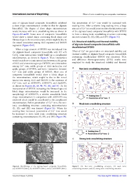
area of alginate-based composite bioscaffolds exhibited The penetration of Ca ions would be increased with
2+
a sheet shape microstructure similar to that for alginate soaking time. After a relative long soaking time, a large
bioscaffold. The degree of sheet shape microstructure amount of Ca ion would penetrate into the microstructure
2+
would increase with ionic crosslinking time as shown in of the alginate-based composite bioscaffolds with SFDDS
Figures 4A and B. Some area of composite bioscaffolds to form a strong ionic crosslinking structure containing
would show a mixed shape containing block shape and microstructures (I), (II), (III), and (IV) (Figure 7C).
fibrous shape in the microstructure, which might be due to
the mixed association among ALG segments and SFDDS 3.3. Structural stability and thermal stability
segments (Figure 4B-F). of alginate-based composite bioscaffolds with
decellularized SFDDS
When a large amount of SFDDS was introduced into
2+
the alginate-based composite bioscaffolds with 0.5 wt% Effect of Ca ion penetration on structural stability and
CaCl , some interactions would build up a complicated thermal stability of alginate-based composite bioscaffolds
2
microstructure, as shown in Figure 6. These interactions containing decellularized SFDDS was discussed. TGA
would contribute to ionic interaction between acidic group and difference thermogravimetry (DTG) results were
of ALG and ammonium group of SFDDS, ionic interaction employed to study the structural stability and thermal
among Ca ions, acidic groups of ALG molecules, and
2+
acidic groups of SFDDS, and ionic interaction between A
Ca ions and acidic groups of SFDDS. Most area of
2+
composite bioscaffolds would show a block shape in
the microstructure, which might be due to the mixed
interaction among ALG and SFDDS in the existence of
Ca ions, such as ADDS1T5, ADDS2T5, and AddS3T5,
2+
as shown in Figures 4E, 4F, 5B, 5C, 6B, and 6C. As the
incorporation of SFDDS increasing, the fibrous shape and
block shape microstructure would be increased. In the
morphology of ADDS3T5, a relative remarkable block
shape microstructure in comparison with ADDS2T5 was
observed which would be contributed to the complicated
microstructure. Before penetration of Ca ions, the non- B
2+
ionic crosslinking structure containing microstructures
(I), (II), and (III) was formed (Figure 7A). When the
soaking time increased, penetration of Ca ions would
2+
be increased to form weak ionic crosslinking structure
containing microstructures (I), (II), and (III) (Figure 7B).
A B
C
C
Figure 6. Morphology of the alginate-based composite bioscaffolds. Figure 7. (A) Non-ionic crosslinking structure, (B) weak ionic
(A) ADDS3T1 (300×), (B) ADDS3T5 (300×), and (C) ADDS3T5 (100×). crosslinking structure, and (C) strong ionic crosslinking structure.
Volume 9 Issue 1 (2023) 41 http://doi.org/10.18063/ijb.v9i1.625