
Page 98 - IJB-9-1
P. 98
International Journal of Bioprinting Progress in bioprinting of bone
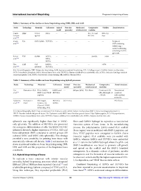
Table 2. Summary of the studies on bone bioprinting using DBB, LBB, and AAB
Study Technology Materials Cell source Animal Pore size Mechanical Compressive Viability Zonal structure
model (porosity) reinforcement modulus
Duarte Inkjet COL-I, MSCs --- --- --- 18.1, 53.1 and 98% at 21 ---
Campos agarose 89.1 kPa days
et al. [130]
Anada SLA OCP, GelMA HUVECs --- --- --- --- --- Peripheral
et al. [131] OCP-containing
GelMA ring +
central GelMA ring
containing HUVEC
spheroids
Bernal VBP GelMA ACPCs, --- --- --- 266 kPa >85% ---
et al. [132] MSCs,
ECFCs
Heo AAB Cell hMSCs, --- --- --- --- >85% ---
et al [133] spheroid HUVECs
SLA: Stereolithography, VBP: Volumetric bioprinting, AAB: Aspiration-assisted bioprinting, COL-I: Collagen type I, GelMA: Gelatin methacrylate,
OCP: Octacalcium phosphate, MSCs: Mesenchymal stem cells, HUVECs: Human umbilical vein endothelial cells, ACPCs: Articular Cartilage-resident
chondroprogenitor cells, ECFCs: Endothelial colony forming cells, hMSCs: Human MSCs
Table 3. Summary of the studies on bone bioprinting using hybrid processes
Study Technology Materials Cell source Animal Pore size Mechanical Compressive Zonal structure
model (porosity) reinforcement modulus
Cui Extrusion+ SLA PLA, GelMA, hMSCs and --- 260 μm (20%) PLA fibers Construct: 0.38 Vascularized
et al. [53] BMP-2 and VEGF HUVECs GPa; Hydrogel: 10 construct
peptides – 30 kPa with capillary
networks
Rukavina Extrusion + Fibrinogen, HUVECs SCID mice --- --- --- PCL frame
et al. [134] DoD Gelatin, HA, ADSCs
Glycerol, VEGF,
bFGF, HAp, PCL
SLA: Stereolithography, DoD: Drop-on-demand, PLA: Polylactic acid, GelMA: Gelatin methacrylate, BMP-2: Bone morphogenetic protein 2,
VEGF: Vascular endothelial growth factor, HA: Hyaluronic acid, bFGF: basic fibroblast growth factor, HAp: Hydroxyapatite, PCL: Polycaprolactone,
hMSCs: Human mesenchymal stem cells, HUVECs: Human umbilical vein endothelial cells, ADSCs: Adipose-derived stem cells
spheroids was significantly higher than that in hMSC- fibers and GelMA hydrogel to reproduce a vascularized
only spheroids. The addition of HUVECs also promoted Haversian system of bone tissue. In the manufacturing
the osteogenic differentiation of cells. The hMSC/HUVEC process, the polydopamine (pDA)-coated PLA scaffold
spheroids showed a higher expression of COL1, ALP, and (bone region) was immobilized with BMP-2 peptides, and
bone sialoprotein (BSP) compared to control groups (2D then, VEGF peptides were conjugated to GelMA chains
cultured hMSC and hMSC-only spheroids). This strategy (vascular region). PLA scaffolds were pre-seeded with
provided a new possibility for printing bone tissue with hMSCs, whereas hMSCs and HUVECs were embedded
anatomically-relevant cell density. Table 2 summarizes the at a 1:1 ratio in the GelMA hydrogel, where the pDA- and
above-mentioned studies on bone bioprinting using DBB, BMP-2-modification was found to promote cell growth
LBB, and AAB and the properties of the bioprinted bone and spread on the scaffold and the BMP-2 benefited
constructs. osteogenesis. In a dynamic culture of perfusion, notable
3.5. Hybrid bioprinting of bone osteogenesis and the formation of vascular networks can
be observed, as indicated by the higher expression of COL-
To replicate a bone construct with interior vascular I, Ca deposition, and VEGF than in static culture.
networks, hybrid bioprinting processes which integrated
EBB and LBB or DBB have been reported. Cui et al. used Combined bioprinting of ADSCs and HUVECs has
[53]
a hybrid bioprinting platform composed of FDM and SLA. also been reported to promote vascularization in bioprinted
Using this technique, they deposited polylactide (PLA) bone tissue [134] . ADSCs underwent osteogenic differentiation
Volume 9 Issue 1 (2023) 90 https://doi.org/10.18063/ijb.v9i1.628