
Page 452 - IJB-9-2
P. 452
International Journal of Bioprinting Fabrication of 3D functional hydrogel for wound dressings
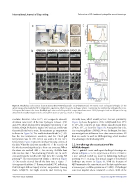
Figure 4. Morphology and structure characterization of the GADQ hydrogels. (a) 3D bioprinter and 3D-printed round and square hydrogels. (b) The
optical images of hydrogel after 3D printing in this experiment. The top row is the hydrogels without crosslinking; the middle is the hydrogels after cross-
linking; and the bottom is the freeze-dried hydrogels after crosslinking. (c) SEM images of the freeze-dried GADQ hydrogels. Scale bar: 500 μm in the top;
200 μm in the middle; 100 μm in the bottom. (d) Pore diameter distributions of the hydrogels.
modulus deviation value (ΔGʹ) and composite viscosity viscosity force, which would perform the best printability.
deviation value (ΔV) of the four hydrogels between 10°C Figure 3g shows the gelation of the GAQ bioink from 25°C
and 25°C, which illustrated the temperature sensitivity of the to 10°C, the complete gel time of four inks decreased from
bioinks. GAQ1.5% had the highest ΔGʹ and ΔV, which was 25°C to 10°C, as shown in Figure 3h. Among the four inks,
theoretically the best to print. The minimum gel temperature the complete gel time of GAQ1.5% was the longest, but there
is shown in Figure 3d. The results indicated that GAQ1.5% was no significant difference from other concentrations. All
has the best temperature sensitivity, and the critical gel four inks could be used for 3D bioprinting, which revealed
temperature was 24.14°C, which was similar to the room the advantages of introducing QCS.
temperature. Figure 3e presents the shear viscosity analysis of
the inks. When the shear rate exceeded 0.1 s , the viscosity of 3.3. Morphology characterization of the
-1
the inks decreased significantly as shear rate increased. When GADQ hydrogels
the shear rate reached 1000 s , the viscosity of all the four The 3D-printed round and square hydrogel dressings are
-1
inks was less than 1 Pa·s, indicating that inks can be easily shown in Figure 4a. In this experiment, a 30 mm × 10 mm ×
extruded from the needles with high shear force during 3D 2 mm cuboid model was used for fabricating hydrogel
printing . The viscoelasticity of bioinks is shown in Figure dressing by 3D printing. The optical images of the printed
[52]
3f. Our results showed that all the inks have a higher Gʹ hydrogels are shown in Figure 4b. With the increase of
(storage modulus) than G˝ (loss modulus) at 25°C, indicating QCS mass ratio, the pore structure of the hydrogels became
that hydrogels did not liquefy at room temperature. Among more compact, and the appearance of GADQ1.5% hydrogel
them, GAQ1.5% had high elasticity and relatively low was more regular when compared to others. SEM of the
Volume 9 Issue 2 (2023) 444 https://doi.org/10.18063/ijb.689