
Page 453 - IJB-9-2
P. 453
International Journal of Bioprinting Fabrication of 3D functional hydrogel for wound dressings
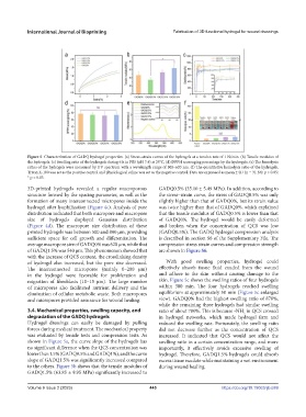
Figure 5. Characterization of GADQ hydrogel properties. (a) Stress–strain curves of the hydrogels at a tension rate of 1 N/min. (b) Tensile modulus of
the hydrogels. (c) Swelling ratio of the hydrogels during 6 h in PBS (pH 7.4) at 25°C. (d) DPPH scavenging percentage by the hydrogels. (e) The hemolysis
ratios of the hydrogels were measured by UV spectrum with a wavelength range of 500–620 nm. (f) The quantitative hemolysis ratio of the hydrogels.
Triton X-100 was set as the positive control and physiological saline was set as the negative control. Data are expressed as mean ± SD (n = 3). NS: p > 0.05;
* p < 0.05.
3D-printed hydrogels revealed a regular macroporous GADQ0.5% (35.10 ± 5.49 MPa). In addition, according to
structure formed by the spacing parameter, as well as the the stress–strain curve, the stress of GADQ0.5% was only
formation of many interconnected micropores inside the slightly higher than that of GADQ0%, but its strain value
hydrogel after lyophilization (Figure 4c). Analysis of pore was twice higher than that of GADQ0%, which explained
distribution indicated that both micropore and macropore that the tensile modulus of GADQ0.5% is lower than that
size of hydrogels displayed Gaussian distribution of GADQ0%. The hydrogel would be easily deformed
(Figure 4d). The macropore size distribution of these and broken when the concentration of QCS was low
printed hydrogels was between 500 and 800 µm, providing (GADQ0.5%). The GADQ hydrogel compression analysis
sufficient space for cell growth and differentiation. The is described in section S6 of the Supplementary File. The
average macropore size of GADQ0% was 620 μm, while that compression stress strain curves and compression strength
of GADQ1.5% was 540 μm. This phenomenon showed that are shown in Figure S6.
with the increase of QCS content, the crosslinking density
of hydrogel also increased, but the pore size decreased. With good swelling properties, hydrogel could
The interconnected micropores (mainly 0–200 μm) effectively absorb tissue fluid exuded from the wound
in the hydrogel were favorable for proliferation and and adhere to the skin without causing damage to the
migration of fibroblasts (10–15 μm). The large number skin. Figure 5c shows the swelling ratios of four hydrogels
of micropores also facilitated nutrient delivery and the within 300 min. The four hydrogels reached swelling
elimination of cellular metabolic waste. Both macropores equilibrium at approximately 50 min (Figure 5c enlarged
and micropores provided assurance for wound healing. view). GADQ0% had the highest swelling ratio of 878%,
while the remaining three hydrogels had similar swelling
3.4. Mechanical properties, swelling capacity, and ratio of about 780%. This is because -NH in QCS crossed
2
degradation of the GADQ hydrogels in hydrogel networks, which made hydrogel firm and
Hydrogel dressings can easily be damaged by pulling reduced the swelling rate. Fortunately, the swelling ratio
forces during medical treatment. The mechanical property did not decrease further as the concentration of QCS
was evaluated by tensile tests and compression tests. As increased. It indicated that QCS would not affect the
shown in Figure 5a, the curve slope of the hydrogels has swelling ratio in a certain concentration range, and more
no significant difference when the QCS concentration was importantly, it effectively avoids excessive swelling of
lower than 1.5% (GADQ0.5% and GADQ1%), and the curve hydrogel. Therefore, GADQ1.5% hydrogels could absorb
slope of GADQ1.5% was significantly increased compared excess tissue exudate while maintaining a wet environment
to the others. Figure 5b shows that the tensile modulus of during wound healing.
GADQ1.5% (63.85 ± 9.95 MPa) significantly increased to
Volume 9 Issue 2 (2023) 445 https://doi.org/10.18063/ijb.689