
Page 197 - IJB-9-3
P. 197
International Journal of Bioprinting 3D-printed vascularized biofunctional scaffold
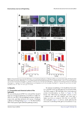
Figure 1. Characterization of each group of hydrogels. (A) 3D printing of each group of hydrogel scaffolds and representative images. (B) Representative
SEM images of each group of hydrogels. (C) X-ray energy dispersive spectral mapping images of PRP-GA@Lap hydrogel: C (red), Si (cyan) and Mg (green).
(D) Pore size of each group of hydrogels (n = 6/group). (E) Porosity size of each group of hydrogels (n = 4/group). (F) Compressive modulus of each group
of hydrogels (n = 4/group). (G, H) The swelling properties and degradation curves of each group of hydrogels (n = 3/group). All experiments were repli-
cated three times, *P < 0.05, **P < 0.01, and ***P < 0.001.
3. Results The internal morphology of the lyophilized GA, PRP-
GA, and PRP-GA@Lap hydrogels was examined by SEM.
3.1. Preparation and characterization of the The SEM images of all hydrogels show interconnected
hydrogels porous microstructures, as shown in Figure 1B. We
We successfully prepared PRP from rats and then mixed it found that PRP-GA@Lap hydrogels tended to have
with GA and Lap in appropriate proportions to make GA, a reduced pore size and rougher pore walls but a
PRP-GA, and PRP-GA@Lap hydrogel precursor solutions, higher porosity as compared with the GA and PRP-
respectively, and then prepared each group of hydrogel GA hydrogels (Figure 1D and E), which indicates the
scaffolds by 3D bioprinting (Figure 1A). We observed that enhanced crosslinking of the gels by the addition of Lap.
PRP-GA@Lap hydrogels had better printing accuracy.
Volume 9 Issue 3 (2023) 189 https://doi.org/10.18063/ijb.702