
Page 268 - IJB-9-3
P. 268
International Journal of Bioprinting Performance of Bredigite-based bone scaffolds
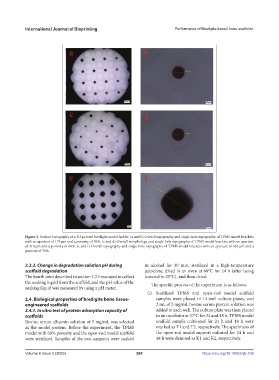
Figure 3. Surface topography of a 3D-printed bredigite model holder. (a and b) Overall topography and single-hole topography of TPMS model brackets
with an aperture of 175 μm and a porosity of 50%. (c and d) Overall morphology and single-hole topography of TPMS model brackets with an aperture
of 310 μm and a porosity of 60%. (e and f) Overall topography and single-hole topography of TPMS model brackets with an aperture of 435 μm and a
porosity of 70%.
2.3.3. Change in degradation solution pH during in alcohol for 30 min, sterilized in a high-temperature
scaffold degradation autoclave, dried in an oven at 60°C for 24 h (after being
The fourth stent described in section 2.3.1 was used to collect lowered to 25°C), and then dried.
the soaking liquid from the scaffold, and the pH value of the The specific process of the experiment is as follows:
soaking liquid was measured by using a pH meter.
(i) Sterilized TPMS and open-rod model scaffold
2.4. Biological properties of bredigite bone tissue- samples were placed in 12-well culture plates, and
engineered scaffolds 2 mL of 5 mg/mL bovine serum protein solution was
2.4.1. In vitro test of protein adsorption capacity of added to each well. The culture plate was then placed
scaffolds in an incubator at 37°C for 24 and 48 h. TPMS model
Bovine serum albumin solution of 5 mg/mL was selected scaffold sample cultivated for 24 h and 48 h were
as the model protein. Before the experiment, the TPMS marked as T1 and T2, respectively. The specimens of
model with 60% porosity and the open-rod model scaffold the open-rod model support cultured for 24 h and
were sterilized. Samples of the two supports were soaked 48 h were denoted as K1 and K2, respectively.
Volume 9 Issue 3 (2023) 260 https://doi.org/10.18063/ijb.708