
Page 91 - IJB-9-3
P. 91
International Journal of Bioprinting Biocompatible 3D printing photosensitive resin
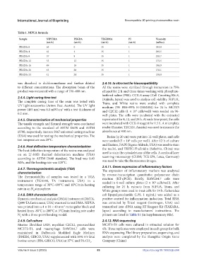
Table 1. NIPUA formula
Sample NIPUMA PEGDA TEGDMA PI Viscosity
(wt.%) (wt.%) (wt.%) (wt.%) (mPa·s)
PEGDA-0 65 0 35 1 183.0
PEGDA-4 61 4 35 1 295.5
PEGDA-8 57 8 35 1 230.7
PEGDA-12 53 12 35 1 175.5
PEGDA-16 49 16 35 1 186.9
PEGDA-20 45 20 35 1 176.1
PEGDA-24 41 24 35 1 138.0
was dissolved in dichloromethane and further diluted 2.4.10. In vitro test for biocompatibility
to different concentrations. The absorption beam of the All the resins were sterilized through immersion in 75%
product was measured with a range of 190–400 nm. ethanol for 2 h and three times washing with phosphate-
buffered saline (PBS). CCK-8 assay (Cell Counting Kit-8,
2.4.4. Light curing time test Dojindo, Japan) was used to analyze cell viability. NIPUA,
The complete curing time of the resin was tested with Trans, and White resins were soaked with complete
UV light accessories (Anton Paar, Austria). The UV light medium (5% FBS+95% H-DEMEM) for 24 h. MC3T3
power (465 nm) was 8.0 mW/cm with a test thickness of and C2C12 cells (1 × 10 cells/well) were seeded on 96-
2
4
0.2 mm. well plates. The cells were incubated with the extracted
2.4.5. Characterization of mechanical properties supernatant for 4, 12, and 24 h. At each time point, the cells
The tensile strength and flexural strength were conducted were incubated with CCK-8 reagent for 1.5 h. A microplate
according to the standard of ASTM D638 and ASTM reader (Sunrise, TECAN, Austria) was used to measure the
D790, respectively. Instron 5967 universal testing machine absorbance at 450 nm.
(USA) was used for testing the mechanical properties. The Resins (φ 20 cm) were put into 12-well plates, and cells
test temperature was 25°C. were seeded (5 × 10 cells per well). After 12 h of culture
4
2.4.6. Heat deflection temperature characterization and fixation, DAPI (Sigma-Aldrich, USA) was used to stain
The heat deflection temperature of the resins was analyzed the nuclei, and TRITC-Phalloidin (Solarbio, China) was
on an LJ-300B thermal deformation machine (USA) used to stain the cytoskeletons of the cells. A confocal laser
according to ASTM D648 standard. The load was 0.45 scanning microscopy (CLSM; TCS SP8, Leica, Germany)
MPa, and the heating rate was 120°C. was used to take the fluorescence images.
2.4.11. Genes expression of inflammatory factors
2.4.7. Thermogravimetric analysis (TGA) The expression of inflammatory markers was analyzed
characterization by reverse-transcription quantitative polymerase chain
The thermostability of samples was tested in a TGA reaction (RT-qPCR). Briefly, RAW264.7 cells were
instrument (TGA550, TA instrument, USA) in a seeded in 6-well culture plates (2 × 10 cells/well). After
5
temperature range of 30°C–600°C and 10°C/min heating culturing for 24 h, extracts from NIPUA, Trans, and
rate in an N atmosphere.
2 White groups were used to treat cells for 24 h. Escherichia
2.4.8. DMA characterization coli lipopolysaccharide (LPS, 1 ng/mL) was added as a
Dynamic mechanical analysis (DMA) instrument (DMTA, positive control for inflammation induction. Total RNA
Q800 TA Instrument, USA) was sued to test DMA. NIPUA was extracted by Trizol reagent (Invitrogen, USA) and
was printed into a 4 × 10 × 40 mm rectangular block and transcribed into cDNA using RT Reagent Kit (TAKARA,
3
heated from -80°C to 200°C at 3°C/min heating rate under Japan) according to manufacturers’ instructions. The
N with a three-point bending model. primers are listed in Table S1 (in Supplementary File).
2
2.4.9. Cell culture 2.4.12. RNA sequencing
Murine fibroblast L929, myoblast C2C12, preosteoblast MC3T3-E1 cells were cultured in extracted solution for
MC3T3-E1, and macrophage RAW264.7 cells were 4 h. Three replicates were employed in each group for bulk
maintained in Dulbecco’s Modified Eagle Medium RNA sequencing. The library preparation, sequencing, and
(DMEM, GIBCO, USA) supplemented with 10% v/v fetal analysis were completed by the ShangHai Origin Gene
bovine serum (FBS, GIBCO, USA) at 37°C and 5% CO . Company (China)
2
Volume 9 Issue 3 (2023) 83 https://doi.org/10.18063/ijb.684