
Page 32 - IJB-9-4
P. 32
International Journal of Bioprinting Lattice-Solid hybrid 3D printing for artificial implant
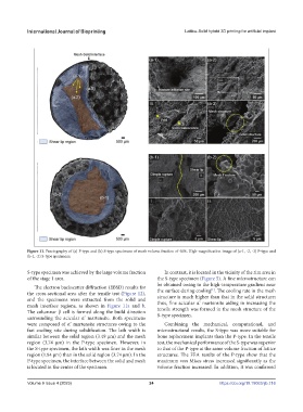
Figure 11. Fractography of (a) P-type and (b) S-type specimens of mesh volume fraction of 40%. High-magnification image of (a-1, -2, -3) P-type and
(b-1, -2) S-type specimens.
S-type specimen was achieved by the large volume fraction In contrast, it is located in the vicinity of the rim area in
of the stage 1 area. the S-type specimen (Figure 5). A fine microstructure can
be obtained owing to the high-temperature gradient near
The electron backscatter diffraction (EBSD) results for [1]
the cross-sectional area after the tensile test (Figure 12), the surface during cooling . The cooling rate in the mesh
structure is much higher than that in the solid structure;
and the specimens were extracted from the solid and thus, fine acicular α’ martensite aiding in increasing the
mesh interface regions, as shown in Figure 11a and b. tensile strength was formed in the mesh structure of the
The columnar β cell is formed along the build direction S-type specimen.
surrounding the acicular α’ martensite. Both specimens
were composed of α’ martensite structures owing to the Combining the mechanical, computational, and
fast cooling rate during solidification. The lath width is microstructural results, the S-type was more suitable for
similar between the solid region (3.49 μm) and the mesh bone replacement implants than the P-type. In the tensile
region (3.74 μm) in the P-type specimen. However, in test, the mechanical performance of the S-type was superior
the S-type specimen, the lath width was finer in the mesh to that of the P-type at the same volume fraction of lattice
region (1.84 μm) than in the solid region (3.74 μm). In the structures. The FEA results of the P-type show that the
P-type specimen, the interface between the solid and mesh maximum von Mises stress increased significantly as the
is located in the center of the specimen. volume fraction increased. In addition, it was confirmed
Volume 9 Issue 4 (2023) 24 https://doi.org/10.18063/ijb.716