
Page 46 - IJB-9-4
P. 46
International Journal of Bioprinting 3D-Bioprinted human lipoaspirate-derived cell-laden skin constructs
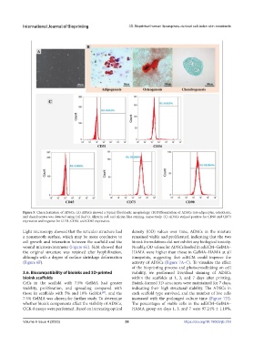
Figure 5. Characterization of ADSCs. (A) ADSCs showed a typical fibroblastic morphology. (B) Differentiation of ADSCs into adipocytes, osteoblasts,
and chondrocytes was detected using Oil Red O, Alizarin red, and Alcian blue staining, respectively. (C) ADSCs stained positive for CD90 and CD73
expression and negative for CD31, CD34, and CD45 expression.
Light microscopy showed that the reticular structure had density (OD) values over time, ADSCs in the mixture
a nonsmooth surface, which may be more conducive to remained viable and proliferated, indicating that the two
cell growth and interaction between the scaffold and the bioink formulations did not exhibit any biological toxicity.
wound microenvironment (Figure 6E). SEM showed that Notably, OD values for ADSCs loaded in adECM–GelMA–
the original structure was retained after lyophilization, HAMA were higher than those in GelMA–HAMA at all
although with a degree of surface shrinkage deformation timepoints, suggesting that adECM could improve the
(Figure 6F). activity of ADSCs (Figure 7A–C). To visualize the effect
of the bioprinting process and photocrosslinking on cell
3.6. Biocompatibility of bioinks and 3D-printed viability, we performed live/dead staining of ADSCs
bioink scaffolds within the scaffolds at 1, 3, and 7 days after printing.
Cells in the scaffold with 7.5% GelMA had greater Bioink-formed 3D structures were maintained for 7 days,
viability, proliferation, and spreading compared with indicating their high structural stability. The ADSCs in
those in scaffolds with 5% and 10% GelMA , and the each scaffold type survived, and the number of live cells
[47]
7.5% GelMA was chosen for further study. To determine increased with the prolonged culture time (Figure 7D).
whether bioink components affect the viability of ADSCs, The percentages of viable cells in the adECM–GelMA–
CCK-8 assays were performed. Based on increasing optical HAMA group on days 1, 3, and 7 were 97.24% ± 1.18%,
Volume 9 Issue 4 (2023) 38 https://doi.org/10.18063/ijb.718