
Page 47 - IJB-9-4
P. 47
International Journal of Bioprinting 3D-Bioprinted human lipoaspirate-derived cell-laden skin constructs
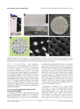
Figure 6. Fabrication of 3D-printed constructs. (A) 3D-bioprinting platform. (B) Bioink was used for printing in a layer-by-layer style in a sterile
environment. (C) Printed constructs exhibited a complex lattice-shaped structure as expected. (D) Image taken by the printer’s high-definition camera. (E)
Representative microscopic image of printed construct. (F) Representative SEM micrograph of printed construct.
95.73% ± 1.21%, and 96.17% ± 1.05%, respectively, of wound closure showed that the wound area decreased
and those in the GelMA–HAMA group were 96.43% ± over time in each treatment group. However, wound sizes
1.07%, 95.35% ± 1.52%, and 95.19% ± 1.4%, respectively. differed among the groups, and on day 7 after implantation,
No significant difference in cell viability was observed the wound area of the blank control group decreased the
between the two bioinks (Figure 7E and F). CCK-8 assay most slowly. On day 14, the two groups with ADSC-laden
and live/dead staining results together demonstrated the scaffolds had achieved complete wound closure. Notably,
biocompatibility of adECM–GelMA–HAMA. We propose the skin in the ADSC-laden adECM–GelMA–HAMA
that the high cell viability in both groups may be related group had healed more completely, whereas that in the
to the nutrient-rich medium used for cell growth in vitro, ADSC-laden GelMA–HAMA group still showed an area
which differs from the complex wound microenvironment of poor epithelization. In both treatments with acellular
in vivo . Although no significant difference was observed scaffolds, a small area of unclosed wound remained, but
[17]
between the two bioink groups, cell viability was >95% in the wound areas in these groups were smaller than those in
both groups. Together, the CCK-8 assay results and live/ the blank control group (Figure 8B).
dead staining results demonstrated the biocompatibility of
both prepared bioinks. Quantitative measurement of wound closure
showed that the area of the unclosed wound at day 7
3.7. In vivo wound healing after treatment with was remarkably smaller in the ADSC-laden adECM–
3D-bioprinted scaffolds GelMA–HAMA group (15.4% ± 1.56%) than that in the
A full-thickness skin defect model in nude mice was used ADSC-laden GelMA–HAMA, adECM–GelMA–HAMA,
to evaluate the effectiveness of ADSC-laden 3D-bioprinted GelMA–HAMA, and blank control groups (26.43% ±
adECM–GelMA–HAMA scaffolds for promoting the 4.11%, 29.47% ± 3.23%, 34.68% ± 3.73%, and 54.48% ±
wound-healing process (Figure 8A). Gross observation 6.59%, respectively). On day 14, the wound area in the
Volume 9 Issue 4 (2023) 39 https://doi.org/10.18063/ijb.718