
Page 48 - IJB-9-4
P. 48
International Journal of Bioprinting 3D-Bioprinted human lipoaspirate-derived cell-laden skin constructs
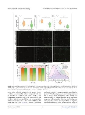
Figure 7. Biocompatibility of bioinks. (A–C) Optical density (OD, at 450 nm) values (CCK-8) of samples of ADSCs cultured in solutions of extract from
adECM–GelMA–HAMA hydrogel for 1, 3, and 5 days. (D) Live/dead staining of cells within printed constructs after 1, 3, and 7 days. Scale bar: 100 μm.
(E) Cell viability. (F) Distribution of cell viability. *p < 0.05, **p < 0.01, and ***p < 0.001.
ADSC-laden adECM–GelMA–HAMA group (0.81% confirmed that ADSCs can accelerate the wound-healing
± 1.14%) was remarkably reduced compared with that process [48-50] . In the early stage of wound healing, implanted
in the adECM–GelMA–HAMA, GelMA–HAMA, and MSCs recruit more endogenous cells through the
blank control groups (6.67% ± 1.97%, 16.65% ± 2.9%, and paracrine effect for tissue remodeling. In the latter stage,
29.42% ± 4.21%, respectively) but was not remarkably exogenous cells gradually disappear, and endogenous
[51]
different from that in the ADSC-laden GelMA–HAMA cells gradually assume the responsibility of repair .
group (0.82% ± 1.52%; Figure 8C). Several studies have Another consideration is that ECM is involved in wound
Volume 9 Issue 4 (2023) 40 https://doi.org/10.18063/ijb.718