
Page 51 - IJB-9-4
P. 51
International Journal of Bioprinting 3D-Bioprinted human lipoaspirate-derived cell-laden skin constructs
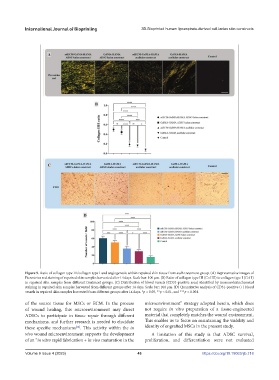
Figure 9. Ratio of collagen type III/collagen type I and angiogenesis within repaired skin tissue from each treatment group. (A) Representative images of
Picrosirius red staining of repaired skin samples harvested after 14 days. Scale bar: 100 μm. (B) Ratio of collagen type III (Col III) to collagen type I (Col I)
in repaired skin samples from different treatment groups. (C) Distribution of blood vessels (CD31-positive area) identified by immunohistochemical
staining in repaired skin samples harvested from different groups after 14 days. Scale bar: 100 μm. (D) Quantitative analysis of CD31-positive (+) blood
vessels in repaired skin samples harvested from different groups after 14 days. *p < 0.05, **p < 0.01, and ***p < 0.001.
of the source tissue for MSCs or ECM. In the process microenvironment” strategy adopted herein, which does
of wound healing, this microenvironment may direct not require in vitro preparation of a tissue-engineered
ADSCs to participate in tissue repair through different material that completely matches the wound environment.
mechanisms, and further research is needed to elucidate This enables us to focus on maintaining the viability and
[64]
these specific mechanisms . This activity within the in identity of engrafted MSCs in the present study.
vivo wound microenvironment supports the development A limitation of this study is that ADSC survival,
of an “in vitro rapid fabrication + in vivo maturation in the proliferation, and differentiation were not evaluated
Volume 9 Issue 4 (2023) 43 https://doi.org/10.18063/ijb.718