Page 398 - IJB-9-5
P. 398
International Journal of Bioprinting                A sturgeon cartilage extracellular matrix-derived bioactive bioink


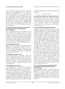
            5 mg, 10 mg, and 15 mg/mLdSC-ECMMA, respectively,   and SerMA hydrogel (Q) can be calculated as the following
            which were correspondingly defined as dSC-ECM-5,   formulation:
            dSC-ECM-10, and dSC-ECM-15. A bioink precursor        Q = (W    – W )/ W  × 100(%)             (I)
            solution containing 150 mg/mLSerMA and 5mg/mL LAP           swollen  dry  dry
            only was prepared as the control bioink sample. Finally,   2.6. Isolation and cultivation of rabbit chondrocytes
            those samples were solidified by exposure to blue light   All animal experiments in this research were approved
            (wavelength: 405 nm) to test their ability to photocuring   by the Animal Care and Ethics Committee of College
            and operability for further cell culture. Hydrogel samples   of Biology, Hunan University. The isolation and culture
            prepared from the dSC-ECMMA containing bioink      of rabbit chondrocytes followed the protocol described
            precursor solutions are simply described as dSC-ECM-5,   in the publication  without  any modification .
                                                                                                           [22]
            dSC-ECM-10, and dSC-ECM-15 hydrogels, respectively,   Chondrocytes in the second passage were harvested for
            in later statements.                               further experiments.
            2.5. Characterizations of hydrogels prepared with   2.7. Cytocompatibility of dSC-ECM hydrogels
            dSC-ECM-derived and the control bioinks            AlamarBlue assays and fluorescein diacetate (FDA)
            2.5.1.  H NMR tests                                staining were carried out to evaluate the cytocompatibility
                 1
            The key component of bioinks, dSC-ECMMA and SerMA,   of prepared hydrogel samples, which encapsulated living
            were characterized and compared with proton nuclear   chondrocytes. SerMA hydrogels, which were prepared
            magnetic resonance ( H NMR) measurements. For the    by photocrosslinking SerMA bioink precursor solution
                              1
            1 H  NMR  analysis,10  mg  of  SerMa  were  completely   carrying chondrocytes (10×10  cells/mL), were used as
                                                                                        5
            dissolved in 1 mL of deuterium oxide (D O; Sigma-Aldrich,   the control samples. The dSC-ECM hydrogel samples
                                           2
            St. Louis, USA). The dSC-ECM and dSC-ECMMA samples   were produced by irradiating dSC-ECM-derived bioinks
            were dissolved as much as possible in D O. All samples in   (10×10  chondrocytes/mL), which contained 5, 10, and
                                                                     5
                                            2
            D O were analyzed by  H NMR (Bruker 400 MHz Advance,   15 mg/mL dSC-ECMMA, respectively (described at 2.4),
                             1
             2
            Switzerland) to obtain a corresponding NMR spectrum.  with blue light. Then, these samples with encapsulated
                                                               chondrocytes were  transferred to  cell culture  plates
            2.5.2. Rheological properties                      with high glucose DMEM containing 10% FBS. The
            Rheological properties of dSC-ECM hydrogels and SerMA   cell culture plates with samples were incubated in a
            hydrogel were evaluatedusing a rheometer (MCR 92,   humidified 37°C, 5% CO  incubator. In brief, the cell-
            Anton Paar GmbH) with oscillation frequency from 1 to   loaded hydrogel samples incubated for 1 and 7 days
                                                                                    2
            10 Hz. The cylinder samples, which were 0.3 mm in height   were taken out, stained with FDA, and observed with
            and 15 mm in diameter cylinder shape, were prepared for   an  inverted  fluorescent  microscope  (IX-73,  Olympus,
            the rheological properties test.                   Japan). The cellular proliferation was analyzed following
                                                                                                      TM
            2.5.3. Mechanical properties tests                 1 and 5 days of incubation using the AlamarBlue  Assay
            The mechanical properties of hydrogel samples were   kit (Thermo Fisher Scientific). Briefly, all samples were
            measured using the electronic universal testing machine   washed three times with PBS and incubated in a fresh
                                                                                                 TM
            (AGS-V universal testing machine with a 20 N sensor,   medium containing 10% v/v AlamarBlue  dye solution
            Shimadzu Corporation). As-prepared cylindrical hydrogel   at 37°C. After 6 h incubation, 200μL of the medium was
            samples (d = 10 mm, h = 3 mm) were placed at the center   collected and measured the fluorescence with excitation at
            of the horizontal measuring platform. All tests were done   570 nm and emission at 630 nm with a spectrophotometer
            on 3 samples (n=3).                                (Thermo Multiskan MK3, USA).

            2.5.4. Equilibrium swelling ratio                  2.8. Scanning electron microscopy and porosity
            The swelling behavior of dSC-ECMMA hydrogels and   measurement of lyophilized dSC-ECM-5 and
            SerMA hydrogels was measured via the gravimetric   SerMA hydrogels
            method. Briefly, samples were immersed for 0.125, 0.5, 1,   The microscopic morphology of samples, which were
            and 1.5 h in 1× PBS (pH 7.4) at 37°C. The hydrogels were   lyophilized dSC-ECM-5 and SerMA hydrogels, were
            washed three times with ddH O, and the filter paper was   observed by scanning electron microscope (SEM) at
                                    2
            used to blot the surface liquid of the hydrogel as much as   an accelerating voltage of 30 kV (FEI Quanta 200, FEI
            possible. The hydrogels were weighed at the different time   Company, Czech Republic). The average pore sizes of the
            points described above to obtain W swollen . Then the gels were   lyophilized hydrogels were analyzed from the SEM images
            lyophilized and measured the dried weight (W ). The   by using ImageJ (50 random pores for each sample were
                                                    dry
            ratio of water uptake from dried dSC-ECMMA hydrogels   calculated).


            Volume 9 Issue 5 (2023)                        390                          https://doi.org/10.18063/ijb.768

