
Page 399 - IJB-9-5
P. 399
International Journal of Bioprinting A sturgeon cartilage extracellular matrix-derived bioactive bioink
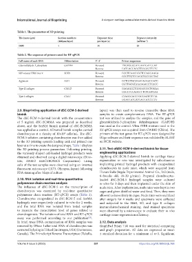
Table 1. The parameters of 3D printing
Thickness (μm) Section numbers Exposure time Exposure intensity
(400μm/layer) per layer (s) (mWcm )
−2
1600 4 30 18
Table 2. The sequence of primers used for RT‐qPCR
Full name of each RNA Abbreviation 5′–3′ Primer sequences
Glyceraldehyde-3-phosphate GAPDH Forward TTGTCGCCATCAATGATCCAT
Reverse GATGACCAGCTTCCCGTTCTC
SRY-related HMG box 9 SOX9 Forward GCGTCAACGGCTCCAGCAAGA
Reverse GCGTTGTGCAGGTGCGGGTAC
Aggrecan AGG Forward GCTGCTACGGAGACAAGGATG
Reverse CGTTGCGTAAAAGACCTCACC
Type II collagen COL II Forward GAGAGCCTGGGACCCCTGGAA
Reverse CGCCTCCAGCCTTCTCGTCAA
Type I collagen COL I Forward CTAGCCACCTGCCAGTCTTTA
Reverse GGACCATCATCACCATCTCTG
2.9. Bioprinting application of dSC-ECM-5 derived Japan) was then used to reverse transcribe these RNA
bioink samples to create complementary DNA. The RT-qPCR
The dSC-ECM-5-derived bioink with the concentration test was utilized to analyze the samples, and the gene of
of 5 mg/mL dSC-ECMMA was prepared as described glyceraldehyde-3-phosphate dehydrogenase (GAPDH)
above, and the SerMA bioink instead of dSC-ECMMA was used as the control. Ultra SYBR mixture used in the
was applied as a control. All tested bioink samples carried RT-qPCR assays was acquired from CWBIO (China). The
6
chondrocytes at a density of 10×10 cells/mL. The dSC- primers of the test genes for RT-qPCR were designed by
ECM-5 solution containing chondrocytes was then added Oligo software and their sequence was validated by BLAST
to the 3D printing system’s loading tank and printed one on NCBI website.
layer at a time to create the designed shape. Table 1 displays
the 3D printing process parameters. Following printing, 2.11. Test ofdSC-ECM-5-derived bioink for tissue
the variously shaped cell-loaded hydrogel products were engineering applications
obtained and observed using a digital microscope (Dino- Applying dSC-ECM-5-derived bioink to cartilage tissue
Lite, ANMO ELECTRONICS Corporation). Living regeneration in vivo was investigated by subcutaneous
cells of the test samples were observed using an inverted implanting printed hydrogel products with encapsulated
fluorescent microscope (IX73, Olympus, Japan) following chondrocytes in nude mice, which were acquired from
FDA staining after 7days of culture. Hunan Slake Jingda Experimental Animal Co., Ltd (male,
6–8weeks old, 18–20 g/mice). Prepared chondrocytes-
2.10. RNA isolation and real-time quantitative loaded dSC-ECM-5 hydrogel samples were cultured
polymerase chain reaction analysis in vitro for 3 days and then implanted under the skin of
The influence of dSC-ECM-5 on the transcription of nude mice. After implantation, nude mice were kept in two
chondrocytes was examined by real-time quantitative cages and given distilled water and food. Then, they were
polymerase chain reaction (RT-qPCR) assays (Table 2). allowed to move freely in cages. Nude mice were sacrificed
Chondrocytes encapsulated in dSC-ECM-5 and SerMA after surgery for 4 weeks and specimens were collected
hydrogels were respectively cultured in vitro for 2 weeks, and subjected to the H&E, SO, and type II collagen
and the total RNA was isolated from tested samples immunohistochemical staining. And stained specimens
to measure the transcription level of genes related to were observed by a microscope to evaluate their in vivo
chondrogenesis. The isolation of total RNA and RT-qPCR cartilage tissue regeneration efficiency.
assay was performed according to our publication .
[22]
In brief, trace DNA contamination of RNA samples was 2.12. Data analysis
removed by DNase I after total RNA of chondrocytes was GraphPad Prism 7.00 was used for statistical computing
extracted by lysing in TRIzol (Invitrogen, USA) (Fermentas, and graph preparation. All data are expressed as mean
Canada). The PrimeScript Reverse Transcriptase (TakaRa, ± standard deviation for a minimum of n=3. Significant
Volume 9 Issue 5 (2023) 391 https://doi.org/10.18063/ijb.768