
Page 403 - IJB-9-5
P. 403
International Journal of Bioprinting A sturgeon cartilage extracellular matrix-derived bioactive bioink
3.4. The microstructure of dSC-ECM-5 hydrogels distributed between 50 and100 μm, and connected pore size
The microstructure of lyophilized dSC-ECM-5 and SerMA of lyophilized SerMA hydrogels was distributed from 30 to
hydrogels were characterized by SEM (Figure 6A). It 60 μm (Figure 6B). It indicated that the connected pores
demonstrated that both lyophilized dSC-ECM-5 and SerMA of dSC-ECM-5 and SerMA hydrogels were available for
hydrogels exhibited a similar porous network structure. effective material transport and signal transmission, which
Pore sizes of lyophilized dSC-ECM-5 hydrogels were mainly are crucial for tissue engineering applications.
3.5. The dSC-ECM-5 bioink for 3D bioprinting
applications
By employing 3D bioprinting, a more advanced 3D
printing technique, hydrogel products with designed
shapes and containing active cells in a suitable
microenvironment can be manufactured and greatly
facilitate tissue engineering applications [25,26] . Strategies
for tissue engineering have been reenergized by the
potential of 3D bioprinting techniques, which not only
achieve the accurate distribution of biomaterials and cells
but also acquire the individual customization of specific
shapes . The projection-based 3D printer was selected as
[27]
a 3D bioprinting machine. The dSC-ECM-5 bioink with
the cell density of 1×10 chondrocytes/mL was used as
6
the row material for 3D printer, and optimized printing
parameters (Table 1) were obtained after three preliminary
trials. Subsequently, the dSC-ECM-5 bioink carrying
chondrocytes was printed into hydrogel products with
designed shapes by using optimized parameters via this 3D
printer (Figure 7). Following bioprinting, it demonstrated
that the produced shapes were high fidelity and clearly
Figure 6. Characterization of dSC-ECM-5and SerMA hydrogel. (A) delineated, suggesting that dSC-ECM-5 is a promising
SEM of lyophilized dSC-ECM-5 and SerMA hydrogel. (B)Pore size
distribution of lyophilized hydrogels. Abbreviations: dSC-ECM-5, 5 mg bioink for 3D bioprinting. Then these hydrogel samples
dSC-ECMMA; SerMa, sericin methacrylate. printed with the dSC-ECM-5 bioink were transferred
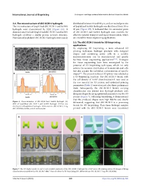
Figure 7. The 3D digital model (A and C) and printed constructs of the dSC-ECM-5 hydrogel by a DLP micro 3D printing system (B).Images of living
chondrocytes encapsulated in dSC-ECM-5 after 7 days of culture by 3D bioprinting (D). Abbreviations: dSC-ECM-5, 5 mg dSC-ECMMA.
Volume 9 Issue 5 (2023) 395 https://doi.org/10.18063/ijb.768