
Page 414 - IJB-9-6
P. 414
International Journal of Bioprinting Biofabrication for islet transplantation
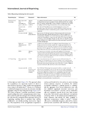
Table 2 Bioprinting technology for islet research
Bioprinting type Cell sources Biomaterial Major achievements Ref
Extrusion-based Mouse pancreatic Alginate, An optimized bioink formulation composed of alginate and gelatin was devel- [89]
bioprinting islets, gelatin oped specifically for the encapsulation of islets and islet-related cells, enabling
endothelial pro- methacryloyl their 3D printing. Additionally, a custom-designed coaxial printer was created
genitor cells (EPC) (GelMA) to facilitate the 3D printing of multicellular constructs that contain islets.
Rat pancreatic dECM Islet-laden dECM bioink was successfully bioprinted to construct 3D [71]
islet, pancreatic tissue. The effectiveness of porcine dECM as a bioink source has
HUVEC, differen- demonstrated its ability to recreate tissue-specific conditions within the 3D
tiated iPSC constructs.
Rat pancreatic islet Alginate, Macroporous 3D hydrogel constructs with precise geometry were success- [88]
methylcellulose fully generated using a plottable hydrogel blend of ultrapure alginate and
methylcellulose (Alg/MC), both of which have been approved clinically. This
enabled the encapsulation of pancreatic islets while preserving their viability,
morphology, and functionality. Notably, the islets within the Alg/MC hydrogel
group maintained their viability and functionality, sustaining the production
of insulin and glucagon.
MIN6-m9, dECM, A hybrid encapsulation system was developed using 3D bioprinting, com- [95]
differentiated H1 PCL bining a macroporous polymer capsule with a stagger-type membrane and
hPSCs assemblable structure, along with a nanoporous dECM hydrogel containing
pancreatic islet-like aggregates. This hybrid system demonstrated biocompati-
bility both in vitro and in vivo, as evidenced by M1 macrophage polarization.
MIN6 Alginate, In a long-term in vivo study, a subcutaneously implanted 3D-bioprinted [111]
PCL construct aimed at preventing islet loss demonstrated promising results. Type
1 diabetes mice implanted with these bioprinted constructs exhibited a signifi-
cant threefold increase in insulin secretion and maintained controlled glucose
levels after 8 weeks of implantation.
DLP bioprinting Mouse pancreatic GelMA Mini encapsulation devices with a groove structure, fabricated using DLP [93]
islets printing, were developed to prevent islet cell leakage. In vivo intraperitoneal
glucose tolerance tests conducted at various time points showcased significant
therapeutic improvement in glycemic control. Even after 15 weeks, a sufficient
amount of viable islet cells were found in the retrieved implant.
Rat pancreatic islet dECM, The HAMA/pECM hydrogel demonstrated its ability to maintain islet cell [98]
hyaluronic acid adhesion and morphology in vitro. In vivo experiments further validated its
methacrylate efficacy, showing that the 3D-printed islet-encapsulated HAMA/pECM hydro-
(HAMA) gel elevated insulin levels in diabetic mice, regulated blood glucose levels
within the normal range for 90 days, and exhibited rapid insulin secretion
in response to blood glucose stimulation. Additionally, the HAMA/pECM
hydrogel promoted the attachment and proliferation of new blood vessels,
leading to an increased density of vascularization.
in blood glucose levels (Figure 8D). This approach allows tool was confirmed both in vitro and in vivo, demonstrating
the encapsulation of a significant number of cells in a reduced immune responses. In addition, the authors
short duration (less than 1 min), thereby averting hypoxic successfully established a printing process to construct
[94]
stress-induced cell dysfunction . Hwang et al. fabricated islet-like aggregates from human pluripotent stem cells
a hybridized encapsulation scheme for pancreatic islet-like that exhibited augmented structural maturation and
[95]
[95]
aggregates using 3D bioprinting technology (Figure 8E) . functional enhancement . Recently, various endothelial
The system comprises a modular macroporous polymer cells and angiogenic growth factors have been printed
capsule configuration with a nanoporous pancreatic tissue- using islets. One research group developed microspheres
derived dECM (pdECM) hydrogel, which effectively shields containing VEGF on 3D-printed poly(dimethylsiloxane)-
the cells from the mechanical forces generated during 3D based constructs and loaded the islets, resulting in
[96]
bioprinting, enabling them to maintain their viability, significant vascularization within 4 weeks . Vascular
proliferation, and insulin-secreting functionality (Figure cells such as endothelial progenitor cells (EPCs), human
8F). Biocompatibility of the amalgamated encapsulation umbilical vein endothelial cells (HUVECs), and induced
Volume 9 Issue 6 (2023) 406 https://doi.org/10.36922/ijb.1024