
Page 415 - IJB-9-6
P. 415
International Journal of Bioprinting Biofabrication for islet transplantation
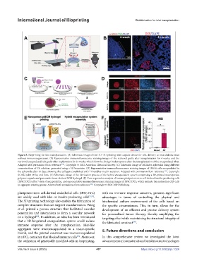
Figure 8. Bioprinting for islet transplantation. (A) Schematic image of the DLP-3D printing mini-capsule device for islet delivery to treat diabetic mice
without immunosuppressant. (B) Representative immunofluorescence staining images of the retrieved grafts after transplantation for 4 weeks, and the
retrieved encapsulated islet grafts after implantation for 15 weeks, which show the foreign body response after the transplantation of the encapsulated islets.
Adapted with permission from reference [93] . Copyright © 2022 American Chemical Society. (C) Schematic image of cell-laden spheroids using different
concentrations of TA solution, generated using a 3D bioprinter. (D) Representative immunofluorescence staining images of INS1E cells encapsulated in
the spheroids after 10 days, showing that collagen crosslinked with TA enables insulin secretion. Adapted with permission from reference [94] . Copyright
© 2022 John Wiley and Sons. (E) Schematic image of the fabrication process of the hybrid encapsulation system comprising a 3D-printed macroporous
polymer capsule and pancreatic tissue-derived ECM hydrogel. (F) Gene expression analysis of human pluripotent stem cell-derived insulin-producing cells
(hPSC-IPCs) after 7 days of encapsulation, and representative immunofluorescence staining images of hPSC-IPCs, which indicate the maturation of β-cells
[95]
in aggregate printing group. Adapted with permission from reference . Copyright © 2021 IOP Publishing.
pluripotent stem cell-derived endothelial cells (iPSC-ECs) with no immune response concerns, presents significant
are widely used with islet or insulin-producing cells [71,97] . advantages in terms of controlling the physical and
The 3D printing technology also enables the fabrication of biochemical culture environment of the cells based on
complex structures that can support vascularization. Wang the specific circumstances. This, in turn, allows for the
et al. printed a porous structure that facilitated vascular development of an efficient and precise delivery system
penetration and interactions to form a vascular network for personalized tumor therapy, thereby amplifying the
on a hydrogel . In addition, an idea has been introduced targeting effect while maintaining the structural integrity of
[98]
that a 3D-bioprinted encapsulation system could reduce the fabricated construct .
[99]
immune response after the transplantation. Islet-like
aggregates were microencapsulated in a tissue-specific 5. Future directions and conclusion
bioink, and the printed construct was macroencapsulated
in a PCL container that blocked immune cells . Moreover, In this comprehensive review, we investigated the latest
[95]
the utilization of genetically modified cells in bioprinting, advancements in biomaterials and biofabrication technologies
Volume 9 Issue 6 (2023) 407 https://doi.org/10.36922/ijb.1024