
Page 409 - IJB-9-6
P. 409
International Journal of Bioprinting Biofabrication for islet transplantation
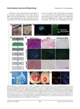
Damodaran and Vermette developed a novel approach of islet functionality within the decellularized pancreata
for perfusion decellularization of the entire pancreas, in vitro, and that the decellularized pancreas liberated
which allows for the engraftment of pancreatic cells and pseudo-islets from the matrix (Figure 4B). Additionally,
islets while maintaining the organ within the perfusion their study highlighted the favorable cytocompatibility
system (Figure 4A). Their work demonstrated the retention of beta-like cells within the decellularized pancreata,
Figure 4. Application of decellularized tissue-driven ECM. (A) Photographic images illustrating the process of pancreas decellularization over a span of
12 h. (B) Immunohistochemistry staining image of GFP-transfected INS-1 cells on a decellularized pancreas, and the image of decellularized pancreas
liberating pseudoislets from the matrix. Adapted with permission from reference . Copyright © 2018 John Wiley and Sons. (C) Schematic image of
[72]
the procedure involved in generating a dECM hydrogel from fragments of human pancreatic parenchyma. (D) H&E staining and immunohistochemistry
staining image of native tissue, decellularized pancreatic hydrogels, and hydrogels for assessing the histological and morphological characteristics. Adapted
with permission from reference . Copyright © 2018 Sackett et al. (E) Photographic image of the process of pancreatic decellularization captured after the
[73]
360-min decellularization procedure. (F) Photographic images of the repopulated pancreatic skeleton enveloped by the omentum for transplantation. (G)
Representative immunofluorescence staining images of iron-oxide-labeled islets in ECM skeletons at 21 and 49 days post-transplantation into the omentum.
Adapted with permission from reference . Copyright © 2022 Berkova et al.
[74]
Volume 9 Issue 6 (2023) 401 https://doi.org/10.36922/ijb.1024