
Page 316 - v11i4
P. 316
International Journal of Bioprinting 3D scaffold prevents tendon ossification
no significant inflammatory hyperplasia or scar tissue, and and molecular signaling (IHC), establishing a robust
preserved tendon morphology (Figure 7C4). framework for evaluating therapeutic outcomes. 62–65
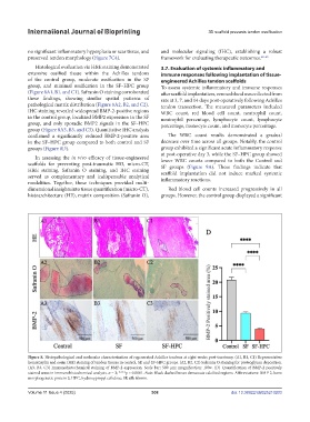
Histological evaluation via H&E staining demonstrated 3.7. Evaluation of systemic inflammatory and
extensive ossified tissue within the Achilles tendons immune responses following implantation of tissue-
of the control group, moderate ossification in the SF engineered Achilles tendon scaffolds
group, and minimal ossification in the SF–HPC group To assess systemic inflammatory and immune responses
(Figure 8A1, B1, and C1). Safranin O staining corroborated after scaffold implantation, venous blood was collected from
these findings, showing similar spatial patterns of rats at 3, 7, and 14 days post-operatively following Achilles
pathological matrix distribution (Figure 8A2, B2, and C2). tendon transection. The measured parameters included
IHC staining revealed widespread BMP-2-positive regions WBC count, red blood cell count, neutrophil count,
in the control group, localized BMP2 expression in the SF neutrophil percentage, lymphocyte count, lymphocyte
group, and only sporadic BMP2 signals in the SF–HPC percentage, monocyte count, and monocyte percentage.
group (Figure 8A3, B3, and C3). Quantitative IHC analysis
confirmed a significantly reduced BMP-2-positive area The WBC count results demonstrated a gradual
in the SF–HPC group compared to both control and SF decrease over time across all groups. Notably, the control
groups (Figure 8D). group exhibited a significant acute inflammatory response
at post-operative day 3, while the SF–HPC group showed
In assessing the in vivo efficacy of tissue-engineered lower WBC counts compared to both the Control and
scaffolds for preventing post-traumatic HO, micro-CT, SF groups (Figure 9A). These findings indicate that
H&E staining, Safranin O staining, and IHC staining scaffold implantation did not induce marked systemic
served as complementary and indispensable analytical inflammatory reactions.
modalities. Together, these techniques provided multi-
dimensional insights into tissue quantification (micro-CT), Red blood cell counts increased progressively in all
histoarchitecture (HE), matrix composition (Safranin O), groups. However, the control group displayed a significant
Figure 8. Histopathological and molecular characterization of regenerated Achilles tendons at eight weeks post-tenotomy. (A1, B1, C1) Representative
hematoxylin and eosin (HE) staining of tendon tissues in control, SF, and SF–HPC groups. (A2, B2, C2) Safranin O staining for proteoglycan deposition.
(A3, B3, C3) Immunohistochemical staining of BMP-2 expression. Scale bar: 500 μm; magnification: 100×. (D) Quantification of BMP-2 positively
stained areas in immunohistochemical analysis. n = 3; ****p < 0.0001. Note: Black dashed boxes demarcate calcified regions. Abbreviations: BMP-2, bone
morphogenetic protein 2; HPC, hydroxypropyl cellulose; SF, silk fibroin.
Volume 11 Issue 4 (2025) 308 doi: 10.36922/IJB025210203