Page 392 - v11i4
P. 392
International Journal of Bioprinting                                      Bioprinted vascular tumor model


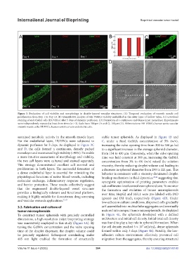
            Figure 3.  Evaluation  of  cell  viability  and  morphology  in  double-layered  vascular  structures.  (A)  Temporal  evaluation  of  smooth  muscle  cell
            proliferation from Day 1 to Day 10. (B) Quantitative analysis of HA-VSMCs viability embedded in the outer layer of hollow tubes. (C) Live/dead
            staining of endothelial cells (HUVECs) after 5 days of dynamic perfusion. (D) Formation of a continuous and dense inner monolayer. Experiments
            were independently repeated at least three times (n ≥ 3). Scale bars: 300 µm (A and C); 100 µm (D). Abbreviations: HA-VSMCs, human aortic vascular
            smooth muscle cells; HUVECs, human umbilical vein endothelial cells.


            sustained metabolic activity in the smooth muscle layer.   stable tumor spheroids. As displayed in  Figure 4B  and
            For the endothelial layer, HUVECs were subjected to   C, under a fixed GelMA concentration of 5% (w/v),
            dynamic perfusion for 5 days. As displayed in Figure 3C   increasing the valve opening time from 200 to 500 μs led
            and D, the cells formed a continuous, densely packed   to a significant increase in the average spheroid diameter,
            monolayer and maintained high viability (>90%). To enable   from 154 to 430 μm. Conversely, when the valve opening
            a more intuitive assessment of morphology and viability,   time was held constant at 300 μs, increasing the GelMA
            the two cell layers were cultured and stained separately.   concentration from 3% to 6% (w/v) raised the solution
            This strategy demonstrated excellent cell survival and   viscosity, thereby reducing droplet volume and leading to
            proliferation in both layers. The successful formation of   a decrease in spheroid diameter from 293 to 221 μm. This
            a dense endothelial layer is essential for mimicking the   behavior is consistent with a viscosity-dominated droplet
            physiological functions of native blood vessels, including   breakup mechanism in fluid dynamics, 45,46  suggesting that
            molecular exchange, inflammatory response  regulation,   synergistic optimization of printing parameters enables
            and barrier protection. These results collectively suggest   sub-millimeter-level control over spheroid size. To monitor
            that the engineered double-layered vessel structure   the formation and evolution of tumor microspheroids
            provides a biologically relevant and functional platform,   over time, HepG2 and HFL1 were pre-labeled with DiO
            making it highly suitable for downstream drug screening   (green)  and  DiI  (red),  respectively  (Figure  4D).  Under
            and vascular research applications. 43,44          low-adhesion culture conditions, dispersed cells gradually

            3.3. Fabrication and culture of                    self-assembled into multicellular aggregates and eventually
            tumor microspheroids                               matured into compact tumor microspheroids. As displayed
            To construct tumor spheroids with precisely controlled   in  Figure 4E, the spheroids developed with a defined
            dimensions, a high-resolution inkjet bioprinting strategy   architecture and initial cell density. Initial total cell density
            was innovatively employed in this study (Figure 4A). By   was found to play a key role in spheroid formation. When
            tuning the  GelMA concentration and the valve opening   the cell density reached 5 × 10⁷ cells/mL, dense spheroids
            time of the droplet dispenser, the droplet volume could   formed within only 3 days (Figure 4E). Notably, the low-
            be precisely regulated. Subsequent crosslinking under   adhesion culture environment effectively prevented cell
            405 nm light enabled the formation of structurally   migration from the aggregates, thereby ensuring structural

            Volume 11 Issue 4 (2025)                       384                            doi: 10.36922/IJB025180180

