
Page 393 - v11i4
P. 393
International Journal of Bioprinting Bioprinted vascular tumor model
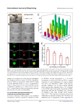
Figure 4. Controlled generation of tumor spheroids via high-precision inkjet bioprinting. (A) Schematic and appearance of the inkjet printing
setup; jetting needle (red-dotted box). (B) Size-controlled tumor spheroids printed by varying valve opening times. (C) Statistical chart of initial
microspheroid diameters prepared under different printing conditions. (D) Fluorescence images of DiO-labeled HepG2 (green) and DiI-labeled HFL1
(red) aggregating into compact spheroids under low-adhesion conditions. (E) Influence of initial total cell density on spheroid formation dynamics.
Experiments were independently repeated at least three times (n ≥ 3). Scale bars: 200 μm (B and D). Abbreviation: GelMA, Methacrylate Gelatin.
stability and consistent size of the spheroids throughout by HUVECs through self-assembly in a 3D ECM)
the subsequent perfusion culture. To further characterize through the co-culture of HUVECs and HFL1. A matrix
spheroid viability, we performed Calcein-AM/PI staining composed of 5 mg/mL fibrinogen and 3 mg/mL collagen
on tumor spheroids at Days 1, 3, and 7. As presented in was used as the extracellular scaffold. Under dynamic
Figure S3, Supporting Information, the results demonstrate perfusion conditions, the co-culture system exhibited
a progressive formation of a central necrotic core by robust microvascular formation (Figure 5A). Compared
Day 7—a typical feature of the tumor spheroid architecture to the HUVEC-only control group, the presence of HFL1
that reflects in vivo solid tumor physiology. significantly enhanced the elongation and maturation
3.4. Construction and characterization of a of endothelial cells, likely through VEGF secretion
multiscale vascularized tumor model (Figure 5B). 47,48 This observation is consistent with previous
To develop a biomimetic TME model, we first established studies demonstrating the synergistic role of fibroblasts and
microvascular networks (prevascular structures formed endothelial cells in angiogenesis, further supporting the
Volume 11 Issue 4 (2025) 385 doi: 10.36922/IJB025180180