
Page 395 - v11i4
P. 395
International Journal of Bioprinting Bioprinted vascular tumor model
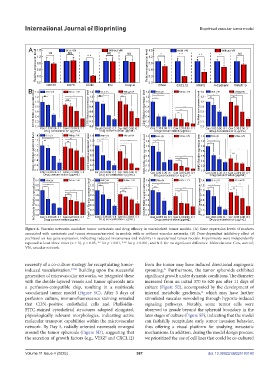
Figure 6. Vascular networks modulate tumor metastasis and drug efficacy in vascularized tumor models. (A) Gene expression levels of markers
associated with metastasis and tumor stemness/survival in models with or without vascular networks. (B) Dose-dependent inhibitory effect of
paclitaxel on key gene expression, indicating reduced invasiveness and viability in vascularized tumor models. Experiments were independently
repeated at least three times (n ≥ 3). p < 0.05, ** for p < 0.01, *** for p < 0.001, and N.S. for no significant difference. Abbreviations: Con, control;
VN, vascular network.
necessity of a co-culture strategy for recapitulating tumor- from the tumor may have induced directional angiogenic
induced vascularization. 49,50 Building upon the successful sprouting. Furthermore, the tumor spheroids exhibited
51
generation of microvascular networks, we integrated these significant growth under dynamic conditions. The diameter
with the double-layered vessels and tumor spheroids into increased from an initial 370 to 620 μm after 11 days of
a perfusion-compatible chip, resulting in a multiscale culture (Figure 5E), accompanied by the development of
vascularized tumor model (Figure 5C). After 3 days of internal metabolic gradients, which may have further
52
perfusion culture, immunofluorescence staining revealed stimulated vascular remodeling through hypoxia-induced
that CD31-positive endothelial cells and Phalloidin- signaling pathways. Notably, some tumor cells were
FITC-stained cytoskeletal structures adopted elongated, observed to invade beyond the spheroid boundary in the
physiologically relevant morphologies, indicating active later stages of culture (Figure 5F), indicating that the model
molecular transport capabilities within the microvascular can faithfully recapitulate early tumor invasion behavior,
network. By Day 5, radially oriented neovessels emerged thus offering a visual platform for studying metastatic
around the tumor spheroids (Figure 5D), suggesting that mechanisms. In addition, during the model design process,
the secretion of growth factors (e.g., VEGF and CXCL12) we prioritized the use of cell lines that could be co-cultured
Volume 11 Issue 4 (2025) 387 doi: 10.36922/IJB025180180