
Page 73 - IMO-2-1
P. 73
Innovative Medicines & Omics Flavonoids against glycosidic hydrolase
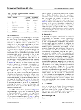
Table 9. Flavonoids in Sophora japonica L. molecular Ser281 residues, the kaempferol-α-glucosidase complex
docking evaluation table showed higher fluctuations than the quercetin-α-
glucosidase complex, indicating that the residue structure
Number Compound α-amylase α-glucosidase
binding energy binding energy was more flexible and unstable. The Rg value of the
(kcal/mol) (kcal/mol) complex stabilized after 50 ns, as shown in the Rg value in
1 Quercetin −5.70 −5.15 Figure 7F. The binding of both quercetin and kaempferol
2 Rutin −3.71 −3.27 increased the Rg value of α-glucosidase. The Rg value of
the α-glucosidase-kaempferol complex was significantly
3 Isorhamnetin −4.54 −4.74 higher than that of the α-glucosidase-quercetin complex,
4 Kaempferol-3-O-rutinoside −3.20 −2.51 which made the whole conformation loose and its structure
5 Kaempferol −4.63 −5.06 unstable, resulting in a decrease in the catalytic activity of
6 Narcissoside −3.14 −2.19 the α-glucosidase active site.
4. Discussion
3.8. MD simulation
In this study, we separated and identified six flavonoids
The binding stability of quercetin/kaempferol-α-amylase from FBSJ and then developed a method to screen their
and α-glucosidase complexes was evaluated using root- inhibitory effects on α-amylase and α-glucosidase activity
mean-square deviation (RMSD), root-mean-square in vitro. The significance of the inhibition was evaluated
fluctuation (RMSF) and radius of gyration (Rg). The through various analytical methods. In addition, we
results of RMSD were used to evaluate the dynamic investigated the inhibitory mechanisms through enzyme
stability of the system. In Figure 7A, the free α-amylase inhibition kinetics, fluorescence spectrometry, CD, and
51
stabilized around 10 ns, and the RMSD of the quercetin- molecular docking. Our analysis indicated that quercetin
α-amylase complex fluctuated in the same region as that and kaempferol were the most effective inhibitors of
of free α-amylase, indicating that quercetin binding α-amylase and α-glucosidase, consistent with the IC
50
only slightly changed the degree of freedom of protein and molecular docking results. Both quercetin and
movement, with stability comparable to that of free kaempferol were found to act as competitive inhibitors
α-amylase. The RMSD value of the kaempferol-α- against α-amylase and α-glucosidase enzymes. The study
amylase complex was significantly higher than that of revealed that quercetin exhibited a mixed inhibition
the quercetin-α-amylase complex, suggesting relatively pattern against α-amylase, while kaempferol demonstrated
poorer stability. The flexibility of amino acid is shown a mixed inhibition pattern against both α-amylase and
in Figure 7B, reflecting the RMSF. During the whole α-glucosidase. Fluorescence and CD spectra revealed
simulation process, the peptide chain from residues 105 that flavonoids in FBSJ caused a reduction in fluorescence
– 111 in the quercetin-α-amylase complex fluctuated intensity and structural changes in the enzymes.
greatly. The combination of kaempferol and α-amylase Molecular docking analysis revealed that quercetin and
caused residues 200 – 213 and 238 to fluctuate greatly. kaempferol exhibited higher binding energies and tighter
These differences in the protein-ligand complexes reflect interactions with the enzymes. MDs simulations provided
the interactions between small molecules and proteins, further insights into the structure and characteristics
which changes the protein conformation and may relate of the α-amylase-quercetin/kaempferol complexes and
to the protein’s functional role. Rg is an important α-glucosidase-quercetin/kaempferol complexes. Notably,
parameter to examine the density of the protein structure; the α-amylase/α-glucosidase-quercetin complexes were
the larger the Rg value of the system, the looser the more stable compared to the α-amylase/α-glucosidase-
protein structure. 52,53 As shown in Figure 7C, the binding kaempferol complexes.
of quercetin reduced the Rg value of α-glucosidase,
indicating that quercetin binds to α-glucosidase and 5. Conclusion
forms a more stable system, thus showing significant Based on scoring for significance, the chromatographic
strong inhibitory activity. analysis CAADA employed in this study proved to be a
The RMSD value of free α-glucosidase in Figure 7D simple and reliable method. Overall, the findings suggest
fluctuated around 0.22 nm, and the RMSD value of the that quercetin and kaempferol exhibit potential as
α-glucosidase-kaempferol complex was significantly inhibitors of α-amylase and α-glucosidase in FBSJ.
higher than that of α-glucosidase-quercetin complex.
All ligand-α-glucosidase complexes in Figure 7E showed Acknowledgment
similar fluctuations. Among the Lys233, His 279, and None.
Volume 2 Issue 1 (2025) 67 doi: 10.36922/imo.6010