
Page 57 - OR-1-2
P. 57
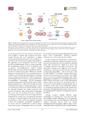
A                                  B


                           C                                  D


            Figure 5. The generation of vascularized brain organoids using induction techniques. (A) Co-culture methods for generating pre-vascularized cerebral
            organoids. (B) Growth factor-induced brain vascularization. (C) Fusion for vascular and brain organoids to achieve vascularization of brain organoids.
            (D) Gene editing for vascularized brain organoids. Image created using BioRender.com.
            Abbreviations: ANG-1: Angiopoietin 1; BOs: Brain organoids; ECs: Endothelial cells; FGF: Fibroblast growth factor; hESCs: Human embryonic stem
            cells; hETV2: Human ETS variant 2; hPSCs: Human pluripotent stem cells; TGF-β: Transforming growth factor-beta; VEGF: Vascular endothelial
            growth factor; VOs: Vascularized organoids; V-Organoids: Vascularized organoids.

            and incorporated them into BOs; however, this approach   The resulting vessel-like structures displayed characteristic
            often struggles to establish fully functional and mature   blood  vessel  ultrastructures,  including  basement
            vascular networks. Shi  et al.  established a co-culture   membranes, EC–cell junctions, and microvesicles.
                                     83
            model by growing human BOs with human umbilical vein   For the development of functionally vascularized BOs,
            endothelial cells (HUVECs) to form tight, embryoid body-  multicellular models that incorporate pericytes, neurons,
            like aggregates, which subsequently underwent neural   astrocytes, and neural stem cells are essential for achieving
            induction and differentiation. In the co-cultured organoids,   robust BBB characteristics.  Studies have shown that
                                                                                     85
            the vascularization process occurred in synchrony with   co-culturing astrocytes with brain microvascular cells
            neurogenesis.  Immunofluorescence  analysis  revealed   significantly increases barrier tightness, while pericytes
            that the major basement membrane glycoprotein laminin   contribute by raising transendothelial electrical resistance
            and the ECs’ marker isolectin B4 co-labeled the vascular   and decreasing permeability – both of which are crucial
            structures, indicating that HUVECs successfully formed a   for BBB integrity. 86,87  In addition, secreted factors from
            complex vascular network within the organoids. Compared   astrocytes and pericytes have been found to induce BBB-
            to non-vascularized organoids, the vascularized organoids   specific properties in endothelial progenitor cells, further
            exhibited significantly increased size and a notably thicker   supporting the establishment of a physiologically relevant
            neuroepithelium.  Interestingly,  HUVEC-vascularized  BBB.  However, challenges remain in fully replicating the
                                                                  88
            organoids demonstrated a reduction in the hypoxia marker   complex cellular interactions and signaling dynamics of
            hypoxia-inducible factor 1-alpha (HIF-1α) and lower levels   in vivo NVUs. Advancements in bioengineering, such as
            of apoptosis, indicating that vascularization plays a critical   microfluidic systems and tailored extracellular matrices,
            role in promoting cell survival. Moreover, HUVECs within   are emerging as promising methods to better simulate these
            the organoids expressed key proteins associated with   interactions, facilitating the development of more accurate
            vascular development, such as P-glycoprotein, suggesting   and vascularized BOs’ models for studying brain function
            that neural cells can modulate ECs’ gene expression,   and disease.
            contributing to the formation of the NVU. Single-cell RNA   Although  co-culture  methods  effectively  induce
            sequencing also revealed that vascularization accelerated   vascularization by integrating ECs or pericytes with BOs,
            neurogenesis, resulting in a higher number of spontaneously   their scalability remains  challenging  due  to inconsistencies
            firing neurons in the vascularized organoids compared   in cell interactions and structural organization. Maintaining
            to their non-vascularized counterparts. Worsdorfer  et   consistent interactions between neural and vascular
            al.  further advanced vascularization in BOs by fusing   components becomes increasingly complex in larger cultures,
              84
            mesodermal precursor cell aggregates with the organoids.   potentially leading to variability in vascular network formation.


            Volume 1 Issue 2 (2025)                         8                                 doi: 10.36922/or.8162