
Page 35 - OR-1-3
P. 35
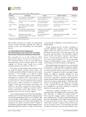
Table 3. Challenges in metabolic studies of brain organoids
             Challenge             Description               Impact             Potential solutions  References
             Oxygen and    Larger organoids → limited diffusion   Core cell death/altered states →   ↓ Organoid size, ↑ media   7,133
             nutrient diffusion  → hypoxia and necrosis in core  skewed metabolic data  optimization, ↑ perfusion systems
             Cellular      Diverse cell types → varying metabolic  Mixed metabolic signals →   Isolate cell populations or design   60,138,139
             heterogeneity  demands                  difficult interpretation  cell type-specific models
             Glucose-induced   Hyperglycemic media → oxidative   Alter metabolic profiles →   Refine glucose levels, monitor ROS   7,140
             stress        stress, ↑ ROS, impaired neuronal   inconsistent outcomes  to reduce stress
                           differentiation
             Enzyme activity  Transcriptomics≠enzyme function →   Enzymatic activity unmeasured  Combine transcriptomics with   7,60,141
                           lack of functional metabolic insight  → incomplete metabolic   enzyme activity assays
                                                     understanding
             Spatial       Imaging mass spectrometry →   Poor spatial resolution of   Develop improved spatial profiling   132,137
             metabolomics  technically complex; hard to integrate   metabolic data  and integration tools.
                           with transcriptomics
             Abbreviation: ROS: Reactive oxygen species.

            these  hurdles,  researchers  can enhance  the  physiological   necessitating the development of faster alternatives such as
            relevance of brain organoids, improving their utility for   voltage imaging.
            studying human brain development and neurological   Voltage imaging provides real-time monitoring of
            diseases.                                         neuronal membrane potential across networks using
            4.3. Advanced functional imaging and              voltage-sensitive dyes (VSDs) and GEVIs. 154,167  VSDs offer
            electrophysiology in brain organoids: Unraveling   high-speed, network-wide activity measurements but can
                                                                                                    168
            neural circuit maturation and disease mechanisms  be invasive and susceptible to dye bleaching,  whereas
                                                              GEVIs provide non-invasive, long-term expression in
            Brain organoids serve as  in vitro models for NDDs,   specific neuronal populations but have slow kinetics
            replicating  key  aspects  of  human  brain  development. 15,52    and limited spatial resolution. 167,169,170  These approaches
            Their functional activity is assessed using patch-clamp   enhance electrophysiological studies, dynamic neuronal
            electrophysiology, calcium imaging, MEAs, and voltage   activity analysis, and network dynamics investigations.
            imaging, each offering unique insights into neuronal
            maturation and disease mechanisms (Table 4). 7,142  MEAs facilitate high-throughput, long-term monitoring
                                                              of extracellular voltage fluctuations, capturing network-
               Patch-clamp electrophysiology remains the gold standard   wide electrical events in real-time. 2D MEAs are effective
                                                                                         171
            for single-cell analysis, providing precise control over   for studying network dynamics but lack the complexity of
            membrane potential and detailed measurements of synaptic   3D systems.  Advances in 3D MEA technology enhance
                                                                        172
            responses. 17,163  This technique can be applied to whole   spatial and  temporal  resolution, allowing for  more
            mount, sliced, or dissociated organoids, each with distinct   comprehensive analysis of neuronal network formation
            advantages and limitations. Whole-mount organoids offer   and activity in organoids.  Integration with optogenetics
                                                                                   151
            a comprehensive view of neuronal activity but are limited   enables targeted neuronal stimulation, improving the study
            to surface neurons,  while sliced and dissociated organoids   of specific cell types in neural circuits.  Traditional MEA
                           143
                                                                                             152
            provide access to deeper structures at the expense of 3D   systems, initially designed for 2D cultures, face challenges
            integrity.  Patch-seq, which integrates patch-clamp with   in fully capturing 3D organoid complexity, prompting
                   146
            scRNA-seq, has further advanced the understanding of   the development of specialized platforms for improved
            molecular mechanisms underlying neuronal activity.    electrode placement and recording.
                                                          164
            However, patch-clamp remains labor-intensive, requires   Fluorescence imaging techniques such as whole-
            specialized training, and has limited throughput.
                                                              organoid and live-cell imaging enable non-invasive, long-
               Calcium imaging enables non-invasive, high-throughput   term observation of cellular and network activity. 157,158
            monitoring of neuronal network activity using genetically   Whole-organoid imaging captures morphological and
            encoded calcium indicators (GECIs) such as GCaMP.  This   functional changes but are limited to surface measurements,
                                                      165
            method provides valuable insights into synaptic plasticity   while live-cell imaging provides high temporal resolution
            and network maturation, particularly when combined with   for tracking dynamic processes such as migration and
            optogenetics for precise neuronal control.  Despite its   differentiation, though it requires advanced microscopy
                                                166
            advantages for long-term studies, calcium imaging has slow   and can cause phototoxicity.  In vivo imaging, particularly
                                                                                    173
            kinetics that limit the resolution of rapid neuronal events,   two-photon microscopy, allows for high-resolution, deep-

            Volume 1 Issue 3 (2025)                         9                            doi: 10.36922/OR025100010