
Page 37 - OR-1-3
P. 37
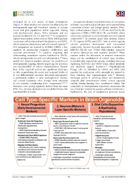
developed for  in vivo studies of brain development   As organoids advance toward maturation, the emergence
            (Figure 3). These markers are essential for identifying the   of distinct neuronal and glial subtypes can be assessed using
            developmental stage and functional identity of various   a repertoire of layer- and lineage-specific markers. Deep-
            neural and glial populations within organoids. During   layer cortical neurons (Layers V–VI) are identified by the
            early developmental phases, NPCs dominate and are   expression of TBR1, CTIP2 (BCL11B), and FOXP2, which
            typically localized in the VZ and SVZ.  VZ progenitors’   collectively contribute to corticothalamic and corticospinal
                                            176
            express transcription factors such as PAX6, which governs   connectivity.  In  contrast,  upper-layer  neurons (Layers
                                                                        182
            neural stem cell identity and early fate decisions, and SOX2,   II–IV) express SATB2 and CUX1, both of which regulate
            which maintains pluripotency and self-renewal capacity.    callosal projection development and intracortical
                                                          177
            SVZ progenitors are marked by EOMES (TBR2), a key   connectivity. General  neuronal maturation is  marked  by
            regulator of intermediate progenitor proliferation and   RBFOX3 (NeuN) and TUBB3 (βIII-tubulin), indicative
            neuronal commitment.  In addition, migrating and   of axonal development and neuronal identity.  These
                                                                                                     182
                                177
            differentiating neuroblasts express doublecortin (DCX),   markers  enable  the  delineation  of  neuronal  maturation
                                                     178
            which is indispensable for early cortical lamination.  These   stages and the establishment of cortical layer identities.
            spatial and temporal markers enhance the resolution of   In parallel, glial populations emerge, including astrocytes
            developmental mapping, thereby improving the precision   expressing ALDH1L1 and GFAP, which reflect metabolic
            and  reproducibility  of  cellular  characterization.  Despite   and structural support functions.  Oligodendrocyte
                                                                                            183
            their utility, organoid systems face significant challenges   lineage cells are identified by OLIG2 and MBP, both
            in fully recapitulating the spatial and temporal fidelity of   essential for myelination, while microglia are marked by
            in vivo differentiation processes. Structural heterogeneity   Iba1, denoting their immunological roles.  However,
                                                                                                  184
            is particularly evident in early neuroepithelial rosettes,   challenges persist in achieving robust and functionally
            and cortical lamination often diverges from canonical   complete glial diversification within current organoid
            in  vivo patterns, complicating direct comparisons. 10,179-181    models, thereby limiting their translational fidelity. The
            Moreover, even among organoids derived from the same   complexity of developmental trajectories necessitates the
            hPSC line, intrinsic developmental variability hinders the   use of multiple markers for accurate cell type identification.
            reproducibility of results.                       Furthermore, the lack of standardized protocols across


                                           Figure 3. Cell type-specific markers in brain organoids


            Volume 1 Issue 3 (2025)                         11                           doi: 10.36922/OR025100010