
Page 38 - OR-1-3
P. 38
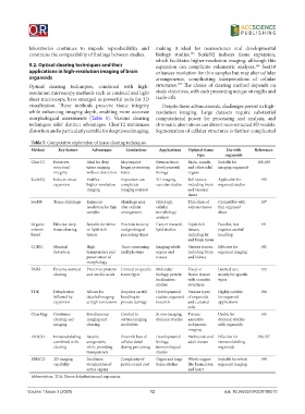
laboratories  continues  to  impede  reproducibility  and   making  it  ideal  for  neuroscience  and  developmental
                                                                           185
            constrains the comparability of findings between studies.  biology studies.  ScaleSQ induces tissue expansion,
                                                              which facilitates higher-resolution imaging, although this
            5.2. Optical clearing techniques and their        expansion can complicate  volumetric  analyses.  SeeDB
                                                                                                     186
            applications in high-resolution imaging of brain   enhances resolution for thin samples but may alter cellular
            organoids                                         arrangements, complicating interpretations of cellular
                                                                      187
            Optical clearing techniques, combined with high-  structures.  The choice of clearing method depends on
            resolution microscopy methods such as confocal and light   study objectives, with each presenting unique strengths and
            sheet microscopy, have emerged as powerful tools for 3D   trade-offs.
            visualization.  These  methods  preserve  tissue  integrity   Despite these advancements, challenges persist in high-
            while enhancing imaging depth, enabling more accurate   resolution imaging. Large datasets require substantial
            morphological assessments (Table 5). Various clearing   computational power for processing and analysis, and
            techniques  offer  distinct  advantages:  ClearT2  minimizes   chromatic aberrations can distort reconstructed 3D models.
            distortion and is particularly suitable for deep tissue imaging,   Segmentation of cellular structures is further complicated

             Table 5. Comparative exploration of tissue clearing techniques
             Method     Key feature  Advantages   Limitations  Applications  Optimal tissue   Use with   References
                                                                              type       organoids
             ClearT2  Preserves    Ideal for deep   May require   Neuroscience,   Brain, muscle,   Suitable for   188,189
                      structural   tissue imaging   longer processing   developmental   and other solid   imaging organoids
                      integrity    without distortion  times  biology     organs
             ScaleSQ  Induces tissue   Enables   Expansion can   3D imaging,   Soft tissues,   Applicable for   190
                      expansion    higher-resolution   complicate   vascular studies  including brain  organoid studies
                                   imaging      imaging analysis          and vascular
                                                                          tissue
             SeeDB    Tissue shrinkage  Enhances   Shrinkage may   Histology,   Thin slices of   Compatible with   187
                                   resolution for thin  alter cellular   cellular   various tissues  thin organoid
                                   samples      arrangement   morphology              slices
                                                              analysis
             Organic   Effective deep   Suitable for dense   Potential toxicity   Cancer research,  Lipid-rich   Possible, but   191
             solvent-   tissue clearing  or lipid-rich   and prolonged   lipid studies  tissues,   requires careful
             based                 tissues      processing times          including fat   handling
                                                                          and brain tissue
             CUBIC    Minimal      High         Time-consuming   Imaging whole   Various tissues,  Effective for   192
                      distortion   transparency and   multiple steps  organs and   including brain  organoid imaging
                                   preservation of            tissues     and kidney
                                   morphology
             PARS     Enzyme-assisted   Preserves proteins  Limited to specific  Molecular   Fixed or   Limited use;   193
                      clearing     and nucleic acids  tissue types  biology, protein   frozen tissues   mostly for specific
                                                              localization   with complex   types
                                                              studies     structures
             TDE      Dehydration   Allows for   Requires careful   Developmental   Various types   Highly suitable   194
                      followed by   detailed imaging   handling to   studies, organoid  of organoids   for organoid
                      expansion    at high resolutions  prevent damage  research  and cultured   applications
                                                                          cells
             ClearMap  Combines    Simultaneous   Limited to   In vivo imaging,   Tissues   Usable for   195
                      clearing and   imaging and   certain imaging   dynamic studies  amenable   dynamic studies
                      imaging      clearing     modalities                to dynamic   with organoids
                                                                          imaging
             iDISCO   Immunolabeling   Retains   Potential loss of   Developmental   Embryonic and  Effective for   196,197
                      combined with   antigenicity   cellular detail   biology,   adult tissues  immunolabeling
                      clearing     while providing   during processing  immunological   organoids
                                   transparency               studies
             3DISCO   3D imaging   Facilitates   Complexity of   Organ and large   Whole organs   Suitable for whole   198
                      capability   visualization of   protocol and cost  tissue studies  like brain, liver,  organoid imaging
                                   entire organs                          and heart
             Abbreviation: TDE: Tissue dehydration and expansion.


            Volume 1 Issue 3 (2025)                         12                           doi: 10.36922/OR025100010