
Page 36 - OR-1-3
P. 36
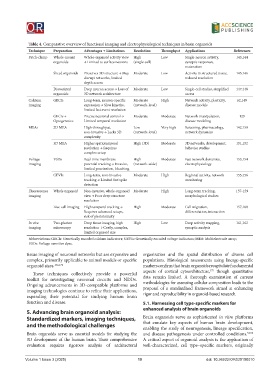
Table 4. Comparative overview of functional imaging and electrophysiological techniques in brain organoids
             Technique  Preparation  Advantages → Limitations  Resolution  Throughput  Applications  References
             Patch-clamp  Whole-mount   Whole-organoid activity view   High   Low  Single-neuron activity,   143,144
                        organoids    → Limited to surface neurons  (single-cell)  synaptic responses,
                                                                                maturation
                        Sliced organoids  Preserves 3D structure → May   Moderate  Low  Activity in structured tissue,   145,146
                                     disrupt networks, limited                  reduced resolution
                                     depth access
                        Dissociated   Deep neuron access → Loss of   Moderate  Low  Single-cell studies, simplified   147,148
                        organoids    3D network architecture                    access
             Calcium    GECIs        Long-term, neuron-specific   Moderate   High  Network activity, plasticity,   62,149
             imaging                 expression → Slow kinetics,   (network-level)  disease models
                                     limited fast event resolution
                        GECIs +      Precise neuronal control →   Moderate  Moderate  Network manipulation,   129
                        Optogenetics  Limited temporal resolution               disease modeling
             MEAs       2D MEA       High-throughput,     Low         Very high  Screening, pharmacology,   142,150
                                     non-invasive → Lacks 3D   (network-level)  network dynamics
                                     complexity
                        3D MEA       Higher spatiotemporal   High (3D)  Moderate  3D networks, development,   151,152
                                     resolution → Requires                      behavior studies
                                     complex setup
             Voltage    VSDs         Real-time membrane   High        Moderate  Fast network dynamics,   153,154
             imaging                 potential tracking → Invasive,   (network-wide)  electrophysiology
                                     limited penetration, bleaching
                        GEVIs        Long-term, non-invasive   Moderate  High   Regional activity, network   155,156
                                     tracking → Limited fast spike              monitoring
                                     detection
             Fluorescence   Whole organoid  Non-invasive, whole-organoid  Moderate  High  Long-term tracking,   157-159
             imaging                 view → Poor deep structure                 morphological studies
                                     resolution
                        Live-cell imaging  High temporal tracking →   High  Moderate  Cell migration,   157,160
                                     Requires advanced setups,                  differentiation, interaction
                                     risk of phototoxicity
             In vivo    Two-photon   Deep tissue imaging, high   High  Low      Deep activity mapping,   161,162
             imaging    microscopy   resolution → Costly, complex,              synaptic analysis
                                     limited organoid size
             Abbreviations: GECIs: Genetically encoded calcium indicators; GEVIs: Genetically encoded voltage indicators; MEA: Multielectrode array;
             VSDs: Voltage-sensitive dyes.

            tissue imaging of neuronal networks but are expensive and   organization and the spatial distribution of diverse cell
            complex, primarily applicable to animal models or specific   populations. Histological assessments using lineage-specific
            organoid sizes. 162,174                           markers confirm that brain organoids recapitulate fundamental
                                                                                          175
               These techniques collectively provide a powerful   aspects of cortical cytoarchitecture,  though quantitative
            toolkit for investigating neuronal circuits and NDDs.   data  remain  limited.  A  thorough  examination  of  current
            Ongoing advancements in 3D-compatible platforms and   methodologies for assessing cellular composition leads to the
            imaging technologies continue to refine their applications,   proposal of a standardized framework aimed at enhancing
            expanding their potential for studying human brain   rigor and reproducibility in organoid-based research.
            function and disease.                             5.1. Harnessing cell type-specific markers for
                                                              enhanced analysis of brain organoids
            5. Advancing brain organoid analysis:
            Standardized markers, imaging techniques,         Brain  organoids serve as  sophisticated  in vitro  platforms
            and the methodological challenges                 that emulate key aspects of human brain development,
                                                              enabling the study of neurogenesis, lineage specification,
            Brain organoids serve as essential models for studying the   and disease pathogenesis under controlled conditions. 52,56
            3D development of the human brain. Their comprehensive   A critical aspect of organoid analysis is the application of
            evaluation  requires  rigorous  analysis  of  architectural   well-characterized, cell type–specific markers, originally


            Volume 1 Issue 3 (2025)                         10                           doi: 10.36922/OR025100010