
Page 14 - GPD-2-2
P. 14
Gene & Protein in Disease MOR in cancer
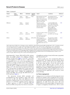
Table 1. (Continued)
Tumors Drugs Effects Mechanism Signaling Result Conclusion References
(Opioids) pathway
NSCLC Tumor Tumor VEGF MOR expression increased The expression level of [130]
promoter angiogenesis significantly in cancer MOR might be associated
samples from patients with with tumor progression
lung cancer compared with
adjacent control tissue
Melanoma Tumor Immune There was a positive µ-opioid peptides might [89]
promoter function correlation between the play a major role in cancer
expression of β-endorphin progression by modulating
and tumor progression in immune response. This
melanoma tissues finding might have
implications for future
optimization of immune-
interventions for cancer
HCC Tumor Tumor MAPK The downregulation of MOR Blocking MOR has [88]
promoter progression inhibited both potential in cancer therapy
in vivo and in vitro human
liver cancer progression
NSCLC Tumor Tumor AKT-mTOR MOR overexpression The exploration of MOR [131]
promoter progression increased AKT and mTOR in NSCLC merits further
activation, proliferation, study both as a diagnostic
and extravasation in human and therapeutic option
bronchioloalveolar carcinoma
cells
AKT: Protein kinase B; B2AR: Beta-2 adrenergic receptor; cAMP/PKA: cyclic adenosine monophosphate/protein kinase A; CRC: Colorectal carcinoma;
EGFR: Epidermal growth factor receptor; ERK: Extracellular signal-regulated kinase; GSK3: Glycogen synthase kinase 3; HCC: Hepatocellular
carcinoma; HIF-1α: Hypoxia-inducible factor 1 alpha; HNSCC: Head and neck squamous cell carcinoma; JAK3/STAT5: Janus kinase 3/signal
transducer and activator of transcription 5; LLC: Lewis lung carcinoma cells; mAb: monoclonal antibody; MAPK: Mitogen-activated protein kinase;
MOR: Mu (µ)-opioid receptor; mTOR: Mammalian target of rapamycin; NADH: Nicotinamide adenine dinucleotide; NFAT: Nuclear factor of activated
T-cells; NSCLC: Non-small cell lung cancer; OPCML: Opioid-binding protein/cell adhesion molecule; PI3k: Phosphoinositide 3 kinase; Src: Rous
sarcoma oncogene cellular homolog; VEGF: Vascular endothelial growth factor
phosphoinositide 3 kinase (PI3k)/AKT/mTOR pathway, cell proliferation and may be involved in tumor progression
which can further promote the malignant biological and chemoresistance .
[89]
behavior of H460 cells (Figure 4). Lu et al. found that Opioids usually exert their corresponding effects by
[87]
the downregulation of MOR was able to inhibit human agonizing MOR. The different and even opposite effects
LC progression both in vivo and in vitro and detected the of opioids in various cancers may be due to factors such
activation of mitogen-activated protein kinase (MAPK)- as cell type, drug, and dose. Overall, the above reports
related signaling pathways while conducting a clinical study have shown that in different types of tumor cell, the
of patients with LC. Similar results have been reported by expression of MOR is closely related to the occurrence and
Zhang et al. who found that the expression of MOR in development of tumors. MOR is anticipated to be used as
[56]
HCC cells and tissue was higher than in non-tumor cells a novel biomarker and therapeutic target for some cancers,
or adjacent tissue and the specific anti-MOR monoclonal necessitating further exploration and research on cancer
antibody (mAb) 3A5C7 inhibited the proliferation of immunotherapy.
HepG2 and Huh7 cells through the MOR-CD147-p53-
MAPK pathway (Figure 5). 3.2. Tumor angiogenesis
The association between MOR expression and Angiogenesis is an essential process for tumorigenesis, tumor
tumorigenesis has been reported in many other types growth, and metastasis, and it is regulated by various factors.
of cancer. In CRC cells, there were differences in the At present, there is a growing interest in the close relationship
expression of MOR in tumor and control tissues and in the between MOR and tumorigenesis and its influence on tumor
activation of cAMP/PKA signaling pathway, suggesting angiogenesis. As solid tumors grow, tumor cells move
a higher expression of MOR in colorectal cancer . further away from their vascular supply. The hypoxic tension
[88]
Moreover, MOR overexpression has shown to increase PC or hypoxia subsequently stimulates tumor cells to secrete
Volume 2 Issue 2 (2023) 8 https://doi.org/10.36922/gpd.282