
Page 341 - IJB-10-1
P. 341
International Journal of Bioprinting 3D-printed bone scaffolds and biofilm formation
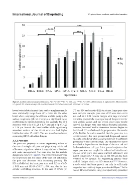
Figure 7. Scaffold surface roughness of Sa and Sq. * for P ≤ 0.05, *** for P ≤ 0.001, and **** for P ≤ 0.0001. Abbreviations: A, high porosity; B, low porosity;
GY, gyroid; RD, reference design; RE, re-entrant auxetic; SD, Schwarz diamond; SP, Schwarz primitive.
lower bacterial attachment, their surface roughness results GY, and SD) and auxetic (RE) structures, larger pore sizes
were statistically insignificant (P > 0.05). On the other were used; for example, pore sizes of SP were 4.04 ± 0.16
hand, when comparing the different scaffold designs, the mm and 3.6 ± 0.01 mm for designs with large and small
surface roughness did not emerge as a significant factor porosities, respectively. A comparison of the pore size for
contributing to biofilm formation. For example, the SP-B each scaffold design and the crystal violet assay results
structure with a Sa of 11.28 ± 1.77 µm and a Sq of 14.23 showed that larger pore sizes reduce bacterial adhesion.
± 2.28 µm formed the least biofilm while significantly However, bacterial biofilm formation became higher in
smoother surface of the RD-B structure had higher the SP and GY scaffolds with larger pore size. The results
biofilm formation (P < 0.05). This was also observed when of the biofilm formation revealed that the pore size is a
comparing RD-B with other designs. specific property for each geometrical design and cannot
be solely considered when deciding between the different
3.3.2. Pore size geometries of the scaffold design. The optimal pore size of
The pore size property in tissue engineering relates to a scaffold is dependent on the shape of the unit cell and
the size of a single cell pore and plays a key role in cell the bacterial/tissue cell type. It is a general conjecture that
adherence, migration, nutrient transportation, infiltration, larger pore sizes are coupled to reduced cell attachment,
and waste management. The pore sizes for the scaffold whereas small pore sizes cause nutrient deficiency and
designs are shown in Figure 8. The pore size is determined inhibit cell growth. A pore size of 100–400 µm has been
50
by the porosity and the shape of the unit cell. Inherently, reported to be optimal for engineering generic bone
the pore size decreases with decreasing porosity. The scaffold designs similar to RD structures. 2,51,52 However,
RD scaffolds had the least pore sizes with RD-A of 0.54 a study stated that bone growth can only be sufficiently
± 0.01 mm and RD-B of 0.17 ± 0.01 mm due to their supported by a relatively larger pore size at 600 µm. In the
53
cellular scaffolding structure. In the case of TPMS (SP, case of TPMS structures, the gyroid specifically presented
Volume 10 Issue 1 (2024) 333 https://doi.org/10.36922/ijb.1768