
Page 419 - IJB-10-1
P. 419
International Journal of Bioprinting Osteogenic differentiation of hMSCs by PBF-LB
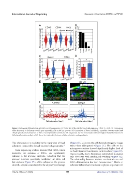
Figure 3. Osteogenic differentiation of hMSCs on 100 μm grooves. (A) Violin plot of the distribution of cells expressing CD90. *p < 0.05. (B) Comparison
of the frequency of the lineage-specific gene-expressing cells on 100 μm grooves. (C) Comparison of Runx2 and PPARγ expression between control and
100 μm grooves. (D) Comparison of the ALP activity between control and 100 μm grooves. (E) The immunocytochemical images of Runx2 expression. (F)
Cellular deformation analysis which shows the relationship between cellular orientation and aspect ratio.
This phenomenon is mediated by the maturation of focal (Figure 3B). Moreover, the cells favored osteogenic lineage
adhesions connected to the cell-secreted collagen matrix. rather than adipogenesis (Figure 3C). The cells on the
23
microgroove surface showed significantly higher level of
Gene sequencing analysis revealed that CD90, which ALP activity rather than those on control surface (Figure 3D).
maintains the stemness of MSCs, was significantly This is possibly due to the nuclear deformation in aligned
reduced in the grooved substrate, indicating that the cells associated with mechanical switching (Figure 3E).
grooved structure genetically facilitated the stem cell The relationship between intrinsic mechanical cues and
fate decision (Figure 3A). MSCs cultured on the grooves MSCs differentiation has been demonstrated. Matrix or
31
showed a specific commitment to the adipose/bone lineage substrate stiffness has been shown to play an important role
Volume 10 Issue 1 (2024) 411 https://doi.org/10.18063/ijb.1425