
Page 418 - IJB-10-1
P. 418
International Journal of Bioprinting Osteogenic differentiation of hMSCs by PBF-LB
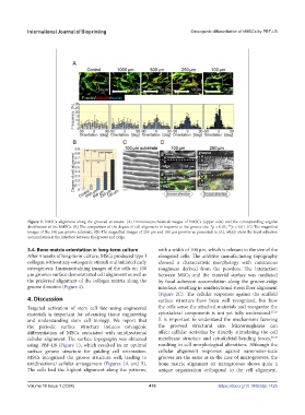
Figure 2. hMSCs alignment along the grooved structure. (A) Immunocytochemical images of hMSCs (upper side) and the corresponding angular
distribution of the hMSCs. (B) The comparison of the degree of cell alignment in response to the groove size. *p < 0.05, **p < 0.01. (C) The magnified
images of the 100 μm groove substrate. (D) The magnified images of 250 μm and 100 μm grooves as presented in (A), which show the focal adhesion
accumulation at the interface between the groove and ridge.
3.4. Bone matrix orientation in long-term culture with a width of 100 µm, which is relevant to the size of the
After 4 weeks of long-term culture, MSCs produced type I elongated cells. The additive manufacturing topography
collagen without any osteogenic stimuli and initiated early showed a characteristic morphology with continuous
osteogenesis. Immunostaining images of the cells on 100 roughness derived from the powders. The interaction
µm grooves surface demonstrated cell alignment as well as between MSCs and the material surface was mediated
the preferred alignment of the collagen matrix along the by focal adhesion accumulation along the groove–ridge
groove direction (Figure 4). interface, resulting in unidirectional stress fiber alignment
(Figure 2C). The cellular responses against the scaffold
4. Discussion surface structure have been well recognized, but how
Targeted activation of stem cell fate using engineered the cells sense the attached materials and reorganize the
materials is important for advancing tissue engineering cytoskeletal components is not yet fully understood. 27,28
and understanding stem cell biology. We report that It is important to understand the mechanisms favoring
the periodic surface structure induces osteogenic the grooved structural size. Microroughness can
differentiation of MSCs associated with unidirectional affect cellular activities by directly stimulating the cell
cellular alignment. The surface topography was obtained membrane structure and cytoskeletal bending forces, 29,30
using PBF-LB (Figure 1), which resulted in an optimal resulting in cell morphological alterations. Although the
surface groove structure for guiding cell orientation. cellular alignment responses against nanometer-scale
MSCs recognized the groove structure well, leading to grooves are the same as in the case of microgrooves, the
unidirectional cellular arrangement (Figures 2A and B). bone matrix alignment on nanogrooves shows quite a
The cells had the highest alignment along the patterns, unique organization orthogonal to the cell alignment.
Volume 10 Issue 1 (2024) 410 https://doi.org/10.18063/ijb.1425