
Page 33 - IJB-2-2
P. 33
Jia Min Lee, Swee Leong Sing, Edgar Yong Sheng Tan, et al.
biomimetic cardiac tissues. Conventional fabrication ratio of ECM proteins that have been isolated in de-
techniques in cardiac tissue engineering can be broad- cellularized matrix are site-specific [47] .
ly divided into scaffold-based or scaffold-free fabrica- Scaffold-based techniques discussed above are ei-
tion. A summary of different conventional fabrication ther laborious or lack repeatability. Firstly, structures
techniques in cardiac tissue engineering is shown in casted out of molds are restricted by the design of
Table 1. master mold. The native structure of myocardium does
(1) Scaffold-based Engineered Cardiovascular not comprise of singular patterns. Instead, cardiomy-
Tissues cytes alignment varies across the transmural of myo-
Scaffolds are used for attachment and mechanical cardium [48] . The effectiveness in using molding app-
support for cardiac cells in scaffold-based approach of oach to recapture complex native architecture is ques-
cardiac tissue engineering. In this approach, cells are tionable.
Another major disadvantage of scaffold-based me-
seeded onto the scaffolds before going through tissue
maturation. In scaffold fabrication, several methods, thod is forming non-uniform macro-pore structure in
such as solvent casting [28–30] , molding [31] and electros- casted scaffolds. An alternative to produce uniform
pinning [32–38] , have been used. Features on the fabri- pore size is to use computer aided technology to de-
cated scaffolds can affect cell responses directly. For sign scaffolds with defined pore structure and size [49] .
example, cellular alignment is shown by neonatal rat Additive manufacturing techniques, such as selective
heart cells seeded on laser microblated polyglycerol laser sintering (SLS) [20] fabricate porous scaffold stru-
sebacate scaffolds that have honeycomb microstruc- ture layer-by-layer with stiffness of fabricated scaffold
ture [39] . similar to native human myocardium (0.2 mPa). Inkjet
In an alternative approach, cells are encapsulated printing technique was also employed for indirect fa-
within the biomaterials. Hydrogel has been widely used brication of scaffolds for tissue engineering [50] .
as the biomaterial to encapsulate cardiac cells [40,41] . Coll- Lastly, perfusion of decellularized matrix, which is
agen ring casted with cardiac cell formed compact heart currently limited to 70% of the original volume, still
muscles and was grafted onto rat’s epicardum [41,42] . To remain a challenge for this scaffolding approach [51] .
improve conductivity of engineered tissue, gold na- Also, decellularized matrix repopulated with neonatal
nowires are mixed with alginate neonatal rat heart rat cardiomyocytes showed disarrayed arrangement
cells and fibroblasts casted into alginate scaffold con- with disorganized electrical propagation and decreased
taining the gold nanowires [43] . Connexion 43 expression [52] . However, decellularized
Another technique scaffold-based technique is to matrix for cardiovascular tissue engineering remains
repopulate de-cellularized matrix with desired cell optimistic with optimization in cell perfusion tech-
population. De-cellularization is a process to obtain niques and improvement on electromechanical proper-
cell-free scaffold from sacrificial tissue/organ through ties of decellularized matrix during the fabrication
removal of xenogenic cells [44,45] . The technique is first process.
used on heart valves and further improvised to repo- (2) Scaffold-free Engineered Cardiovascular Tissues
pulate a full heart [46] . A distinct advantage of de-cellul- As the name suggests, scaffold-free approach does
rized matrix over hydrogel-based scaffold is retaining not make use of solid scaffolds, usually made from
the native extracellular matrix. The composition and polylactic acid (PLA), polycaprolactone (PCL) and etc.,
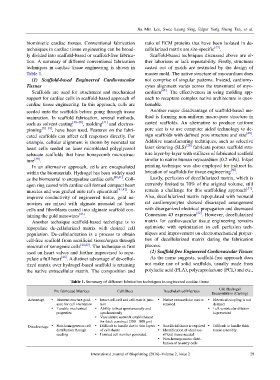
Table 1. Summary of different fabrication techniques in engineered cardiac tissue
Pre-fabricated Matrices Cell Sheet Decellularized Matrices Cell Hydrogel
Encapsulation (Casting)
Advantage Inherent structure guid- Intact cell-cell and cell-matrix junc- Native extracellular matrix Electrical coupling is not
ance for cell orientation tion retained delayed
Tunable mechanical Ability to beat spontaneously and Left ventricular dilation
properties synchronously is prevented
Vasculature network establishment
for thick construct (300 – 800 µm)
Disadvantage Non-homogeneous cell Difficult to handle due to thin layers Sacrificial tissue is required Difficult to handle thick
distribution through of cell sheets Identification of ideal sac- tissue assembly
seeding Limited cell number generated rificial tissue needed
Non-homogeneous distri-
bution of re-entry cells
International Journal of Bioprinting (2016)–Volume 2, Issue 2 29