
Page 468 - IJB-10-2
P. 468
International Journal of Bioprinting Bioprinting with ASCs and bioactive glass
1% 100× antibiotic/antimycotic. On the second day of time to test viscosity, recovery, yield strength, etc. Three
culture, ASCs were washed with phosphate-buffered saline different tests were conducted on the gels: (i) viscosity
(PBS), viable ASCs were harvested with 0.25% trypsin/1 vs. shear rate (to measure viscosity with increasing shear
mM ethylenediaminetetraacetic acid (EDTA), and re- rate from 0.1 to 100 s ), (ii) oscillation amplitude sweep,
-1
plated at a concentration of no more than 15,000 ASCs and (iii) recovery tests (changing shear rate from steady
per dish. ASCs that reached ≤70% confluency were lifted state to a predetermined rate for a certain amount of
between the second and sixth passages for suspension in time). In oscillation amplitude sweeps, percentage strain
AG hydrogels for all experiments. ASCs from subsequent was considered input, and machine output data of loss vs.
passages were not utilized for experiments as they could storage modulus components were plotted. Data points
affect pluripotent properties of ASCs. below 0.1 s shear rate were not reported because of the
-1
instability at low shear rates. Although one set of data was
2.2. Bioink preparation reported for each gel type, measurements were repeated
Gelatin (Type B, Sigma-Aldrich, St. Louis, MO, USA) to confirm the validity of the data. Statistical analysis of
in 3 w/v % (0.3 g in 10 mL) was dissolved in Dulbecco’s the rheological data was not performed because of the
Modified Eagle Medium (DMEM; Gibco, Thermo Fisher significant differences between the samples prepared with
Scientific, MA, USA) in a glass beaker at ~40°C while being several orders of magnitudes difference in results.
magnetically stirred at 150 rpm. After gelatin dissolution,
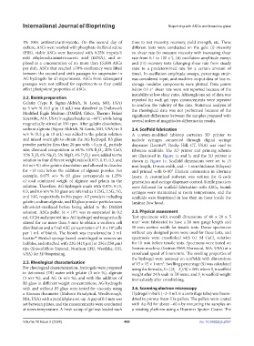
sodium alginate (Sigma-Aldrich, St. Louis, MO, USA) in 3 2.4. Scaffold fabrication
w/v % (0.3 g in 10 mL) was added to the gelatin solution A custom-modified tabletop cartesian 3D printer to
and mixed overnight to obtain the AG hydrogel. B3 glass include syringes connected through digital syringe
powder particles (less than 20 µm with ~3 µm d particle dispenser (Loctite , Rocky Hill, CT, USA) was used to
®
50
size; chemical composition in wt.%: 53% B O , 20% CaO, fabricate scaffolds. The 3D printer and printing schema
2
3
12% K O, 6% Na O, 5% MgO, 4% P O ) were added to the are illustrated in Figure 1a and b, and the 3D printer is
2
2
5
2
solution in four different weight ratios (0.075, 0.15, 0.3, and shown in Figure 1c. Scaffold dimensions were set to 15
0.6 w/v %) after gelatin dissolution and allowed to dissolve mm length, 15 mm width, and ~ 1 mm thickness (6 layers)
for ~10 min before the addition of alginate powder. For and printed with 0–90° filament orientation in alternate
example, 0.075 w/v % B3 glass corresponds to 1.25% layers. A customized software was written for G-code
of total combined weight of alginate and gelatin in the generation and syringe dispenser control. Sterile practices
solution. Therefore, AG hydrogels made with 0.075, 0.15, were followed for scaffold fabrication with ASCs, bioink
0.3, and 0.6 w/v % B3 glass are referred as 1.25G, 2.5G, 5G, syringes were maintained at room temperature, and the
and 10G, respectively, in this paper. All powders including scaffolds were bioprinted in less than an hour inside the
gelatin, sodium alginate, and B3 glass powder particles were laminar flow hood.
ultraviolet-sterilized before being added to the DMEM
6
solution. ASCs pellet (4 × 10 ) was re-suspended in 0.2 2.5. Physical assessment
mL CCM and pipetted into AG hydrogel and magnetically Test specimens with overall dimensions of 40 × 20 × 5
3
stirred for no more than 3 min to obtain a uniform cell mm were fabricated to have a 20 mm gauge length and
distribution and a final ASC concentration of 1.0 × 10 cells 10 mm section width for tensile tests. Dense specimens
6
per 1 mL of bioink. The bioink was transferred to 3 mL without any designed pores were used for these tests, and
Loctite Henkel syringe barrel, centrifuged to remove air specimens were crosslinked with 0.1 M CaCl solution
®
2
bubbles, and attached with 22G (410 µm) or 25G (250 µm) for 10 min before tensile tests. Specimens were tested on
tips (SmoothFlow Tapered, Nordson EFD, Westlake, OH, Instron machine (Instron 5969, Norwood, MA, USA) at a
USA) for 3D bioprinting. crosshead speed of 5 mm/min. The swelling properties of
the hydrogel were assessed on scaffolds with dimensions
2.3. Rheological characterization of 15 × 15 × 1 mm . Swelling percentage (S) was calculated
3
For rheological characterization, hydrogels were prepared using the formula, S = [(S – S )/S] × 100, where S is scaffold
t
c
t
in deionized (DI) water with gelatin (3 w/v %), alginate weight after 24 h soak in DI water, and S is scaffold weight
(3 w/v %), and AG (6 w/v %), and with the addition of immediately after crosslinking. c
B3 glass in different weight concentrations. AG hydrogels
with and without B3 glass were tested for viscosity using 2.6. Scanning electron microscopy
a Kinexus rheometer (Malvern Panalytical, Westborough, Hydrogel in bulk (~2–3 mL in a centrifuge tube) was freeze-
MA, USA) with a parallel plate set-up. A gap of 0.5 mm was dried to porous foam-like pellets. The pellets were coated
set between plates, and the measurements were conducted with Au-Pd for about ~60 s by mounting the samples on
at room temperature. A fresh scoop of gel was loaded each a rotating platform using a Hummer Sputter Coater. The
Volume 10 Issue 2 (2024) 460 doi. 10.36922/ijb.2057