
Page 469 - IJB-10-2
P. 469
International Journal of Bioprinting Bioprinting with ASCs and bioactive glass
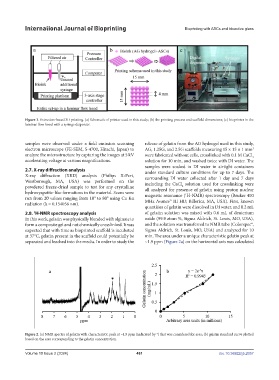
Figure 1. Extrusion-based 3D printing. (a) Schematic of printer used in this study; (b) the printing process and scaffold dimensions; (c) bioprinter in the
laminar flow hood with a syringe dispenser.
samples were observed under a field emission scanning release of gelatin from the AG hydrogel used in this study,
electron microscope (FE-SEM, S-4700, Hitachi, Japan) to AG, 1.25G, and 2.5G scaffolds measuring 15 × 15 × 1 mm
3
analyze the microstructure by capturing the images at 5 kV were fabricated without cells, crosslinked with 0.1 M CaCl
2
accelerating voltage at various magnifications. solution for 10 min, and washed twice with DI water. The
samples were soaked in DI water in airtight containers
2.7. X-ray diffraction analysis under standard culture conditions for up to 7 days. The
X-ray diffraction (XRD) analysis (Philips X-Pert, surrounding DI water collected after 1 day and 7 days
Westborough, MA, USA) was performed on the including the CaCl solution used for crosslinking were
powdered freeze-dried sample to test for any crystalline all analyzed for presence of gelatin using proton nuclear
2
hydroxyapatite-like formations in the material. Scans were 1
run from 2θ values ranging from 10° to 80° using Cu Kα magnetic resonance ( H-NMR) spectroscopy (Bruker 400
MHz Avance™ III HD, Billerica, MA, USA). First, known
radiation (λ = 0.154056 nm).
quantities of gelatin were dissolved in DI water, and 0.2 mL
1
2.8. H-NMR spectroscopy analysis of gelatin solution was mixed with 0.6 mL of deuterium
In this work, gelatin was physically blended with alginate to oxide (99.9 atom %, Sigma Aldrich, St. Louis, MO, USA),
form a composite gel and not chemically crosslinked. It was and the solution was transferred to NMR tube (Colorspec®,
expected that with time as bioprinted scaffold is incubated Sigma Aldrich, St. Louis, MO, USA) and analyzed for 10
at 37°C, gelatin present in the scaffold could potentially be min. The area under a unique characteristic gelatin peak at
separated and leached into the media. In order to study the ~1.9 ppm (Figure 2a) on the horizontal axis was calculated
Figure 2. (a) NMR spectra of gelatin with characteristic peak at ~1.9 ppm (indicated by *) that was considered for area; (b) gelatin standard curve plotted
based on the area corresponding to the gelatin concentration.
Volume 10 Issue 2 (2024) 461 doi. 10.36922/ijb.2057