
Page 478 - IJB-10-2
P. 478
International Journal of Bioprinting Bioprinting with ASCs and bioactive glass
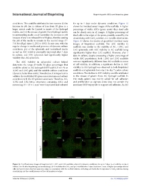
conditions. This could be attributed to two reasons: (i) the for up to 7 days under dynamic conditions. Figure 12
increase in pH due to release of ions from B3 glass to a shows the live/dead assay images of the scaffolds. A higher
larger extent could be limited to inside of the hydrogel percentage of viable ASCs (green spots) than dead cells
matrix, and (ii) the release of gelatin from hydrogel matrix can be clearly seen in all images. A higher percentage of
to surrounding media could neutralize the increase in pH dead cells in the edges of the pores, possibly caused by the
because of any ions released from B3 glass, thereby causing crosslinking with CaCl solution, is a notable observation.
2
the pH of the media to remain in the neutral range (7– Figure 13 shows the results of quantified live/dead assay
7.6) for all gel types (1.25G to 10G). In any case, with the images of bioprinted scaffolds. The ASC viability in
regular change in media and presence of dynamic culture scaffolds was similar to the viability of AG, 1.25G, and
conditions, pH of the spheroids and neutralized media 2.5G spheroids, with ASC viability in AG scaffold being
as well as ASC viability eventually improved after 7 days significantly higher than 2.5G scaffold. However, after 7
in culture, and 2.5G spheroids had significantly higher days in culture, despite possessing a higher percentage of
viability than AG spheroids. viable ASC populations, both 1.25G and 2.5G scaffolds
The ASC viability in spheroidal culture helped were not significantly different than AG scaffolds in terms
determine the range of viable B3 glass percentages that of cell viability. In addition, a significant decline in ASC
could be added to AG hydrogel (0.075 and 0.15 w/v % or viability in AG hydrogel was observed in both bioprinted
1.25G and 2.5G gels) and the suitable culture conditions scaffolds and spheroids from day 1 to day 7 under culture
(dynamic better than static). Nonetheless, it is important to conditions. The decline in ASC viability could be attributed
validate the established B3 glass concentrations and culture to the release of gelatin from AG hydrogel scaffold. In
conditions with the 3D-printed constructs. Therefore, AG, this study, gelatin was mainly added for cell adhesion
1.25G and 2.5G lattice structures containing ASCs and and proliferation as alginate alone does not contain the
3
measuring 15 × 15 × 1 mm were bioprinted and cultured necessary RGD tripeptide to support cell adhesion. In AG
Figure 12. Live/Dead assay images of bioprinted AG, 1.25G, and 2.5G scaffolds cultured in dynamic conditions for up to 7 days. (a–c) viability on day 0
(within 2 to 4 h after bioprinting and crosslinking), (d–f) viability after 24 h, and (g–i) viability after 7 days. Scale bars: 1 mm. A higher percentage of dead
cells (red spots) were observed near pore edges in comparison with the scaffold interior, indicating cell death due to exposure to CaCl solution during
2
crosslinking and the overall presence of low cell numbers at edges.
Volume 10 Issue 2 (2024) 470 doi. 10.36922/ijb.2057