
Page 11 - IJB-4-2
P. 11
Lee J M, et al.
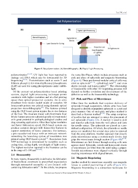
Figure 5. Vat polymerization: (A) Stereolithography. (B) Digital Light Processing.
polymerization [63,64] . UV light has been reported to the name Bio-Placer, which include processes such as
damage cell DNA which may be detrimental for 3D pick and place of spheroids and magnetic bioprinting
bioprinting [65,66] . Photoinitiators such as eosin Y and (Figure 6). These pre-formed modular units of cells may
lithium phenyl-2,4,6-trimethylbenzoylphosphinate exist as spheroids [69–71] , cylindrical rods [70] , and even
(LAP) are used for curing photopolymers under visible- sheets (toroids and honeycombs) [72] . The relationship
light. of bioassembly with other 3D bioprinting proceses will
Of the current vat polymerization-based printing depend on further evolution and development of the
system, digital light processing technique prints definition as well as the bioassembly technology.
constructs with higher resolution and at a fast printing 2.5 Pick and Place of Microtissues
speed than optical projection systems. An in vitro
triculture liver tissue model made of complex 3D Other than the methods that requires delivery of
honeycomb pattern was printed using dynamic optical spheroids through suspensions, robotic arms have been
projection stereolithography [67] . The features printed designed to directly manipulates spheroids in a pick and
using the DLP-based system mimics the in vivo features place manner. One of such pick and place method is
of the liver. The triculture model with biomimetic liver described as Kenzan method [73,74] . The setup comprises
lobule features presents a physiologically relevant model of needles that are arranged in arrays for placement of
with great potential in pathophysiological studies and cell spheroids (Figure 6A). A suction is used to pick
drug screening applications. Other than higher resolution and transfer spheroids from the well plates and onto
and faster fabrication speed, DLP-based system can the needle arrays. The spheroids are placed in a pre-
produce complex designs with lumen-like features to defined configuration. Of which, the spheroids on needle
support maturation of tissue construct. For instance, arrays are incubated for several days prior to removal
a pre-vascularized tissue with an intricate network from the array platform. Another approach that directly
mimicking the branching system of vasculature was picks microtissues using a custom-made device that
printed using DLP-based system [61] . Resolution from grips the microtissues through suction (Figure 6B) [72,75] .
VPP technology is determined by variables such as Microtissues are first formed through seeding cells onto
curing time, curing depth, wavelength of light source. agarose mold. Spheroids, toroids and honeycomb sheets
The highest resolution reported in the literatures can be of microtissues are lifted from the mold using a gripper.
in the range of 5–100 µm [60,61,68] . Toroids microtissues were aligned vertically through
2.4 Bioassembly stacking microtissues with aligned lumens.
In many reports, bioassembly is defined as the fabrication 2.6 Magnetic Bioprinting
of hierarchical constructs in prescribed organization Another method for microtissue assembly uses magnetic
through automated assembly of pre-formed cell- forces to control positioning of cells (Figure 6C).
containing fabrication untis . In this article, we propose Magnetic bioprinting is a contactless technique for
[1]
International Journal of Bioprinting (2018)–Volume 4, Issue 2 5