
Page 12 - IJB-4-2
P. 12
3D bioprinting processes: A perspective on classification and terminology
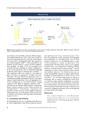
Figure 6. Direct manipulation of cells or modular units of cells: (A) Pick and place spheroids with needles. (B) Pick and place spheroids
using a custom-made device. (C) Pick and place using magnetic forces.
manipulating and assembling cells into different shapes. and spanning across micro- and macro-scales. There
Two distinct methods are used in this setup. Firstly, in have been numerous works in making 3D bioprinting
label-free diamagnetophoretic printing, cell-medium more adoptable for real applications. One of these
was mixed with a paramagnetic buffer and exposed to examples include the use of multimaterials to create
an external magnetic field [76] . Cells suspended in the 3D microchannels to enable vascularization. This
medium moved towards a region of lower magnetic multimaterials 3D bioprinting system can be applied
field strength. The shape of 3D cell assemblies was similarly like a fused deposition modelling (FDM),
controlled through changing the magnet configuration. which is a material extrusion AM technique . Hybrid
[80]
In the second approach, cells are magnetized through bioprinting is another future trend that combine natural
incubating with magnetic nanoparticles overnight [77,78] . and synthetic materials. This hybrid system can use
The magnetized cells were seeded in a low-adherent strong biodegradable polymer as support and bioactive
plate, forming cell aggregates through levitation. hydrogel as model materials to create exterior of 3D
Thereafter, the magnetized cell aggregates were re- scaffolds [51] . As 3D bioprinting advances and more
suspended in medium and patterned using a ring-shaped techniques start to emerge, standardized classification
magnet. Spatial patterning of the cell aggregates into of technology using consistent terminology is necessary
desired morphology are controlled through varying to serve as a baseline towards development of standards
variation in the shape of magnetic template used [79] .
Limitation in the magnetic field strength in constructing for 3D Bioprinting. The proposed classification here
larger construct requires further improvisation for will also promote knowledge and helps to stimulate new
miniaturization [76] . Nevertheless, cytotoxicity and research by defining the processes based on the physical
plau sible internal stresses of the engulfed magnetic principles of the technologies.
nanoparticles may have detrimental effects on the cells. References
A summary of bioprinting and bioassembly technologies
is given in Table 1. 1. Groll J, Boland T, Blunk T, et al., 2016, Biofabrication:
3. Conclusions and Outlook Reappraising the definition of an evolving field.
3D Bioprinting has become an enabling fabrication tool Biofabrication, 8(1): 013001. http://dx.doi.org/10.1088/1758
5090/8/1/013001
in various applications using different material systems
6 International Journal of Bioprinting (2018)–Volume 4, Issue 2