
Page 114 - IJB-5-2
P. 114
Preparation and printability of ultrashort self-assembling peptide nanoparticles
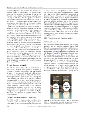
is comprised peptides with no more than 7 amino acid to PBS). Gelation of both peptides occurred within a
residues, capable of self-assembly into supramolecular few minutes at a minimum concentration of 4 mg/mL
fibrous network structures due to their peptide motifs. and 3 mg/mL for CH-01 and CH-02, respectively, as
Through a microfluidics flow-focusing method , we shown in Figure 1. As described in section 2.3, the 3D
[19]
can prepare NPs from ultrashort peptides of different printing system prints using a higher concentration
sequences for applications ranging from drug delivery to of peptide solution as the increased viscosity enables
bioimaging [20,21] . This fabrication method is particularly printing at a higher resolution. Due to this, to prepare
advantageous due to its ability to continually produce the manual hydrogel samples for these experiments, a
peptide NPs at a scale that allows for use in experiments. concentration of 10 mg/mL of peptide was used to ensure
In the past, our laboratory has reported on the use of a final concentration comparable to those of the printed
peptide hydrogels as scaffolds for tissue engineering and samples. For manual sample conditions made with NPs,
regenerative medicine, as well as on the preparation of approximately 0.9 mg of NPs were added to the peptide
hydrogels with slow-releasing silver NPs (AgNPs) for solution before the addition of the PBS either by volume
antimicrobial applications [22-25] . The addition of peptide from the product of the microfluidic chip or in the form
NPs to peptide hydrogels allows for the localized delivery of lyophilized NPs.
of any drugs or growth factors conjugated to the surface 2.2 NP Fabrication and Characterization
of the NPs. This is facilitated by way of a composite of
sorts made entirely from a single material. In addition,
we have published on our novel three-dimensional (3D) 2.2.1 NP Fabrication Process
printer setup where we have explored the printability NPs were fabricated through a microfluidic flow-focusing
of bioinks produced in the laboratory in conjunction method by way of a Dolomite 6 Junction Droplet Chip.
with various cell types [26-30] . Inspired by the potential of This chip has six separate junctions that combine into one
peptide NPs and 3D bioprinting, we decided to combine output channel for increased product. At the junction, the
the two technologies to study the printability of our NPs. main channel is intersected perpendicularly by the two
Two sequences of self-assembling peptides are tested side channels (Figure 2A). The peptide solution in water
and assessed for shape fidelity. The promising results flowing through the main channel is funneled by two
indicate that different to the manual approach the 3D side channels containing 50% (v/v) ethanol solution into
printing of ultrashort self-assembling peptide NPs may a jet-like stream. The pressure from the side channels,
result in hydrogels embedded with a more homogenous through which 50% of ethanol in water solution (v/v)
distribution of NPs. is running focuses the mainstream and leads to NP
formation. Through the flow-focusing mechanism, the
2. Materials and Methods peptide aggregates in the water. The ultrashort peptide
of a given sequence was dissolved in Milli-Q water
The NPs are fabricated through a microfluidic-driven and loaded into a 1 mL syringe to be pushed through
flow-focusing method. The system is comprised a the central channel in the junctions of the chip, and an
Dolomite 6 Junction microfluidic chip (dimensions:
45 mm × 15 mm, channel depth and width at cross-
section: 50 µm × 55 µm), Nikon Eclipse TS 100 inverted
microscope, Harvard Apparatus PhD Ultra syringe pump,
Chemyx Fusion 200 syringe pump, and plastic syringes
(BD, Luer Lok in 10 mL and 1 mL). About 50% (v/v) of
ethanol solution was prepared by diluting absolute ethanol
(Sigma-Aldrich) and then filtering through a Millex-GP
syringe filter with a pore size of 0.22 µm. Tetrameric
self-assembling peptides CH-01 and CH-02 were custom
synthesized in our laboratory for nanomedicine through
solid-phase peptide synthesize and purified to higher
than 95% using preparative high-performance liquid
chromatography.
2.1 Manual Hydrogel Sample Preparation
The CH-01 and CH-02 peptide powders were dissolved in Figure 1. The self-assembling peptides CH-01 (4 mg/ml) and
Milli-Q water, then mixed with ×10 phosphate-buffered CH-02 (3 mg/ml) produce hydrogels in aqueous solution; the
saline (PBS) at a final volume ratio of 9:1 (peptide solution gelation was enhanced using phosphate-buffered saline.
110 International Journal of Bioprinting (2019)–Volume 5, Issue 2