
Page 248 - IJB-10-6
P. 248
International Journal of Bioprinting DIW of concave hydroxyapatite scaffolds
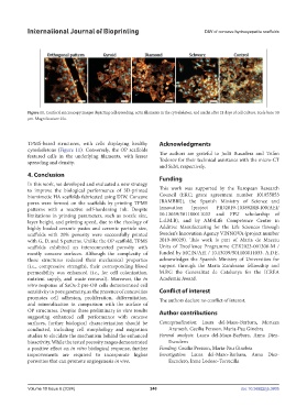
Figure 11. Confocal microscopy images depicting cell spreading, actin filaments in the cytoskeleton, and nuclei after 21 days of cell culture. Scale bars: 50
μm. Magnification: 25×.
TPMS-based structures, with cells displaying healthy Acknowledgments
cytoskeletons (Figure 11). Conversely, the OP scaffolds
featured cells in the underlying filaments, with lesser The authors are grateful to Judit Buxadera and Trifon
spreading and density. Todorov for their technical assistance with the micro-CT
and SEM, respectively.
4. Conclusion
Funding
In this work, we developed and evaluated a new strategy
to improve the biological performance of 3D-printed This work was supported by the European Research
biomimetic HA scaffolds fabricated using DIW. Concave Council (ERC; grant agreement number 101055053
pores were formed on the scaffolds by printing TPMS [BAMBBI]), the Spanish Ministry of Science and
patterns with a reactive self-hardening ink. Despite Innovation (project PID2019-103892RB-I00/AEI/
limitations in printing parameters, such as nozzle size, 10.13039/501100011033 and FPU scholarship of
layer height, and printing speed, due to the rheology of L.d.M.B), and by AM4Life Competence Centre in
highly loaded ceramic pastes and ceramic particle size, Additive Manufacturing for the Life Sciences through
scaffolds with 20% porosity were successfully printed Sweden’s Innovation Agency VINNOVA (project number
with G, D, and S patterns. Unlike the OP scaffold, TPMS 2019-00029). This work is part of Maria de Maeztu
scaffolds exhibited an interconnected porosity with Units of Excellence Programme CEX2023-001300-M /
mostly concave surfaces. Although the complexity of funded by MCIN/AEI / 10.13039/501100011033. A.D.E.
these structures reduced their mechanical properties acknowledges the Spanish Ministry of Universities for
(i.e., compressive strength), their corresponding blood support through the Maria Zambrano fellowship and
permeability was enhanced (i.e., for cell colonization, M.P.G the Generalitat de Catalunya for the ICREA
nutrient supply, and waste removal). Moreover, the in Academia Award.
vitro response of SaOs-2 pre-OB cells demonstrated cell
sensitivity to pore geometry, as the presence of concavities Conflict of interest
promotes cell adhesion, proliferation, differentiation, The authors declare no conflict of interest.
and mineralization in comparison with the surface of
OP structures. Despite these preliminary in vitro results Author contributions
suggesting enhanced cell performance with concave
surfaces, further biological characterization should be Conceptualization: Laura del-Mazo-Barbara, Morteza
conducted, including cell morphology and migration Aramesh, Cecilia Persson, Maria-Pau Ginebra
studies to elucidate the mechanism behind the enhanced Formal analysis: Laura del-Mazo-Barbara, Anna Diez-
bioactivity. While the tested porosity ranges demonstrated Escudero
a positive effect on in vitro biological response, further Funding: Cecilia Persson, Maria-Pau Ginebra
improvements are required to incorporate higher Investigation: Laura del-Mazo-Barbara, Anna Diez-
porosities that can promote angiogenesis in vivo. Escudero, Irene Lodoso-Torrecilla
Volume 10 Issue 6 (2024) 240 doi: 10.36922/ijb.3805