
Page 245 - IJB-10-6
P. 245
International Journal of Bioprinting DIW of concave hydroxyapatite scaffolds
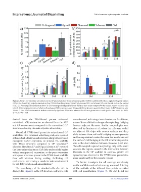
Figure 7. SaOs-2 pre-osteoblast cell cultures on the 3D-printed calcium-deficient hydroxyapatite (CDHA) scaffolds with either a typical orthogonal pattern
(OP) or the three triply periodic minimal surface (TPMS)-based structures (gyroid [G], diamond [D], and Schwarz [S]), and the well plate as the control
(Ctrl). (a) Percentage of cell adhesion after 6 h. (b) Percentage of cell proliferation at different time points compared to the control sample on day 1 (d1).
(c) Cell differentiation, in terms of alkaline phosphatase (ALP) expression, over 21 days. (d) Mineralization quantified by Alizarin Red (AR) staining after
14 and 21 days. (Different numbers indicate statistically significant differences between the different geometries at each specific time point; p < 0.05).
*p < 0.05, **p < 0.01.
derived from the TPMS-based pattern enhanced were observed, indicating a low adhesion rate. In addition,
osteoblastic (OB) maturation, as observed from the ALP most of these cells had an elongated morphology, bridging
and AR measurements, compared to the conventional OP between adjacent filaments. Similar morphologies were
structure, achieving the main objective of our study. observed by Graziano et al., where the cells were seeded
Overall, all TPMS-based geometries outperformed OP on adjacent HA chips with convex surfaces and deep
scaffolds in vitro, consistent with Zhang et al., who reported clefts between them, with cells bridging between granules
enhanced cell adherence and retention, along with increased and having minimal contact between the membrane and
8
osteogenic marker expression, on sintered HA scaffolds the surface. Cell bridging in the OP structure is possible
with TPMS structures compared to OP structures. due to the short distance between filaments (~60 µm).
28
Likewise, Bianchi et al. and Urquia Edreira et al. reported The cells adopted a spread morphology only in the small
71
72
that bone mineralization on CaP disks preferentially begins concave-like regions created in the intersection between
within hemispherical concavities, as the pore concavities filaments in the OP scaffold. In contrast, greater cell
and layered structures present in the TPMS-based scaffolds spreading was observed in the TPMS-based geometries,
favor cell retention during seeding, facilitating cell more significantly in the concave regions.
confinement, and creating a conducive microenvironment To further investigate the cell coverage and density
for cell differentiation and mineralization. on the scaffolds, confocal microscopy was used. Stitching
8
The morphology of the attached cells after 6 h is of the scaffolds at the different time points correlated
displayed in Figure 8. In the OP structure, only a few cells with cell quantification (Figure 9). On day 1, full cell
Volume 10 Issue 6 (2024) 237 doi: 10.36922/ijb.3805