
Page 134 - IJB-8-1
P. 134
Compression Failure of Trabecular Tantalum Scaffolds
Figure 11. Scanning electron microscopy micrographs of the ductile fracture surface of additive manufacturing-fabricated Ta sample after
tensile fracture failure.
A B C
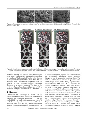
Figure 12. Optical microscope photographs and electron backscattered diffraction micrographs of the metallographic specimens of trabecular
Ta scaffolds with porosity of 65%: (A) Uncompressed original sample. (B) Annealed compressive. (C) unannealed compressive samples.
gradually transmit load through their interconnecting as-fabricated specimens exhibited fully interconnecting
struts to the overall structure, rather than bearing the load and stochastically distributed porous structures
in a local area. The longitudinal (parallel to the direction (Figures 4 and 5) resembling cancellous bone. The
of compressive load) struts bear more stress than the struts had notably dense and smooth appearance after
transverse struts. Under the same compressive strain, the sandblasting, indicating that the starting Ta powders were
stress on the struts of trabecular Ta scaffolds gradually fully fused during LPBF-based AM. The microstructural
decreases as the porosity increases. The stress on the examination (Figure 5) revealed dense struts in the AM-
struts of trabecular Ta scaffolds with porosity of 65% is fabricated trabecular Ta scaffolds after sandblasting. No
the largest among the scaffolds with the 3 porosities. obvious processing defects or fractures were found on the
struts. However, an uneven secondary morphology with
4. Discussion slight irregularities was observed on the surfaces of Ta
LPBF-based AM technology is suitable for the struts. Although these imperfections are unfavorable to
fabrication of porous metallic scaffolds due to its high the mechanical properties of trabecular Ta scaffolds, they
precision and processing repeatability [7,37,38] . In this may be beneficial to improvement of osteoblast adhesion,
study, LPBF was employed to manufacture porous Ta migration, and proliferation. The measurement results of
scaffolds consisting of trabecular structures with various the gravimetric method indicate that the porosities of AM-
porosities (65%, 75%, and 85%) and an identical strut fabricated trabecular Ta scaffolds were approximately
diameter (250 μm). Morphological analysis revealed that 2% smaller than those of the designed models (Table 1),
120 International Journal of Bioprinting (2022)–Volume 8, Issue 1