
Page 135 - IJB-8-1
P. 135
Yang, et al.
A B C
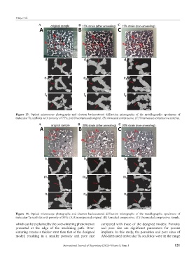
Figure 13. Optical microscope photographs and electron backscattered diffraction micrographs of the metallographic specimens of
trabecular Ta scaffolds with porosity of 75%; (A) Uncompressed original. (B) Annealed compressive. (C) Unannealed compressive samples.
A B C
Figure 14. Optical microscope photographs and electron backscattered diffraction micrographs of the metallographic specimens of
trabecular Ta scaffolds with porosity of 85%: (A) Uncompressed original. (B) Annealed compressive. (C) Unannealed compressive sample.
which can be explained by the over-sintering phenomenon compared with those of the designed models. Porosity
presented at the edge of the machining path. Over- and pore size are significant parameters for porous
sintering creates a thicker strut than that of the designed implants. In this study, the porosities and pore sizes of
model, resulting in a smaller porosity and pore size AM-fabricated trabecular Ta scaffolds were in the range
International Journal of Bioprinting (2022)–Volume 8, Issue 1 121